Stages of Periodontal Disease: What Stage I–IV and Grade A–C Mean
Meet the Stom Dental Centre team
“Generalised periodontitis, stage III, grade B.” A diagnosis like this is precise for the dentist who wrote it and opaque for the person it describes. It comes from the classification that the American Academy of Periodontology and the European Federation of Periodontology agreed at a workshop in Chicago in November 2017 and published in June 2018, which is why you will see both years attached to it.
The system gives two answers. The stage, I to IV, says how much damage there is and how difficult it will be to manage. The grade, A to C, says how fast the disease has been moving and how likely it is to go on. This article sets out the exact criteria for both, shows how the diagnosis is reached, and explains what stage and grade mean for treatment and for keeping your teeth. One thing it cannot do is stage you. The classification is built on measurements with a probe and on X-rays, not on symptoms, and a single stage is given to the whole mouth by a clinician.
- Two parts. Stage (I–IV) describes severity and complexity. Grade (A–C) describes the rate of progression and the risk of more.
- The stage follows the worst-affected tooth. Attachment loss of 1–2 mm between the teeth is stage I, 3–4 mm stage II, 5 mm or more stage III or IV.
- Stage III and IV share the same attachment and bone loss. What separates them is tooth loss (up to 4 teeth, or 5 and more) and complications such as loose, drifting teeth or a collapsing bite.
- Smoking and diabetes move the grade. Ten or more cigarettes a day, or an HbA1c of 7.0% or more in a person with diabetes, mean grade C.
- The stage does not go down after treatment; the grade can change in either direction. Successful treatment makes periodontitis stable. It does not turn the patient into someone who never had it.
- Aftercare is step 4, and it does not end. In studies of patients in maintenance for five years or more, the average loss was about one tooth per patient every ten years, and most patients lost none.
Before the Stages: Health, Gingivitis, Periodontitis
The classification starts with a definition of health, for the first time (Dietrich 2019). Gums count as clinically healthy when fewer than 10% of the measured sites bleed on gentle probing and no supporting tissue has been lost. Gingivitis is bleeding at 10% of sites or more, still without loss of support: localised when 10–30% of sites bleed, generalised above 30% (Sanz 2020, summarising the workshop).
Gingivitis is inflammation confined to the gum, and it is reversible when the bacterial film is removed (Murakami 2018). Periodontitis begins where the attachment of the tooth is lost. A patient counts as a periodontitis case when attachment loss between the teeth can be measured on at least two teeth that are not neighbours, or when at least two teeth show 3 mm or more of attachment loss on the cheek or tongue side together with pockets deeper than 3 mm, and nothing else explains the loss, such as a root fracture or recession caused by injury (EFP guidance notes, 2019).
Does gingivitis always turn into periodontitis? No. In a group of Sri Lankan tea workers who had no dental care and did not clean their teeth, followed from 1970 to 1985 (480 men at the start, 161 at the last examination), about 11% never progressed beyond gingivitis, about 81% progressed moderately and about 8% rapidly (Löe 1986). But inflamed gums are the main warning. In a 26-year study of 565 Norwegian men with regular dental care, 99.5% of teeth with consistently non-inflamed gums were still present at a tooth age of about 50 years, against 63.4% of the few teeth (103) whose gums bled at every examination (Schätzle 2004; Lang 2009).
The disease usually does not hurt. The British Society of Periodontology calls it the silent disease in its patient leaflet and lists the signs people do notice: bleeding when brushing or eating, red and swollen gums, bad breath, gaps appearing between teeth, teeth that loosen or move, receding gums. Our article on gingivitis and periodontitis covers symptoms and causes in detail.
How the Diagnosis Is Made
The European guideline describes four steps for a new patient: suspect periodontitis, confirm it, stage it, grade it (Sanz 2020).
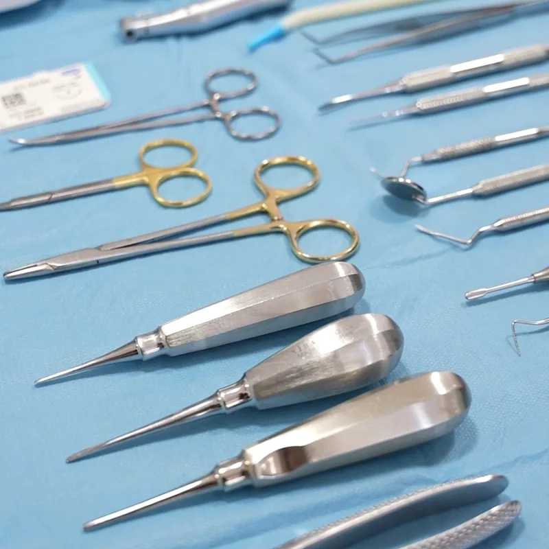
The main instrument is a thin probe with millimetre markings. It measures two things at six points around every tooth. Probing depth is the distance from the gum edge to the bottom of the pocket. Clinical attachment loss is measured from a fixed point on the tooth, the border between crown and root, and therefore also records support that was lost where the gum has receded. The dentist notes which sites bleed. X-rays show how much bone is left in relation to the length of the root. According to the European Federation of Periodontology’s patient page, two small images are enough in simple cases, and extensive cases may need up to 14 more or a panoramic image.
Many patients first meet a screening score, not a stage. In the United Kingdom this is the Basic Periodontal Examination (BPE), which divides the mouth into six sections and records the worst score in each.
| BPE code | What the probe finds | What follows (British Society of Periodontology, 2019) |
|---|---|---|
| 0 | Pockets under 3.5 mm, no bleeding, no tartar or overhanging filling edges | No periodontal treatment needed; screening again at the next routine examination |
| 1 | Pockets under 3.5 mm, bleeding on probing | Oral hygiene instruction; screening again at the next routine examination |
| 2 | Pockets under 3.5 mm, tartar or overhanging filling edges | Oral hygiene instruction and removal of tartar and overhanging edges; screening again at the next routine examination |
| 3 | Probing depth 3.5–5.5 mm | Initial treatment, then full six-point measurements in that section |
| 4 | Probing depth over 5.5 mm | Full six-point measurements of all teeth |
| * | The division between the roots of a back tooth is affected | Recorded in addition to the code |
The society is explicit that the BPE is for screening only and should not be used for diagnosis. Germany uses a similar index, the PSI, with codes 0 to 4 for six sections; people with statutory health insurance are entitled to it every two years, and a suspicious result is followed by a full clinical and X-ray examination (DG PARO). A screening code of 3 or 4 is therefore a reason for a more detailed examination. It is not a stage.
The Four Stages: The Exact Criteria
The table gives the criteria of the American Academy of Periodontology’s sheet in plain language; they come from the framework paper by Tonetti, Greenwell and Kornman (2018).
| Criterion | Stage I | Stage II | Stage III | Stage IV |
|---|---|---|---|---|
| Attachment loss between the teeth, at the worst site | 1–2 mm | 3–4 mm | 5 mm or more | 5 mm or more |
| Bone loss on the X-ray | The third of the root nearest the crown, under 15% | The third of the root nearest the crown, 15–33% | Reaching the middle third of the root or beyond | Reaching the middle third of the root or beyond |
| Teeth lost to periodontitis | None | None | 4 or fewer | 5 or more |
| Local complexity | Probing depth 4 mm at most; mostly even (horizontal) bone loss | Probing depth 5 mm at most; mostly even bone loss | Probing depths of 6 mm or more; vertical bone loss of 3 mm or more; the area between the roots of back teeth clearly affected (class II or III); moderate defects of the jaw ridge | In addition: need for complex rehabilitation because of chewing problems, loose teeth (mobility degree 2 or more), severe ridge defects, bite collapse, drifting or flaring teeth, or fewer than 20 remaining teeth (10 opposing pairs) |
The European Federation of Periodontology gives the stages names: initial (I), moderate (II), severe with potential for additional tooth loss (III), and advanced with extensive tooth loss and potential for loss of the dentition (IV).
Five rules explain how the table is used.
- Attachment loss comes first. If it has not been measured, bone loss on the X-ray is used. Tooth loss from periodontitis can change the stage.
- The worst tooth decides. One stage is given per patient, based on the most affected tooth.
- Complexity can only raise the stage. In general a single complexity factor is enough. Clear involvement of the area between the roots (class II or III) puts a case in stage III or IV whatever the attachment loss.
- Stage III and IV differ mainly in complexity. The guideline’s example: very loose teeth or a collapsed bite at the back point to stage IV.
- Extent is added as a word. “Localised” means fewer than 30% of the teeth are involved, “generalised” means 30% or more; a third pattern affects mainly molars and incisors.

The authors of the system admit its limits. Telling stage I or II apart from stage III or IV is described as relatively uncomplicated, while the finer distinctions need careful judgment by an experienced clinician, and probe measurements carry an error that makes some misclassification of early cases inevitable (Kornman 2020; EFP guidance notes).
A word on stage I. In the sixth German Oral Health Study, which applied the 2018 classification, 31.6% of adults aged 35 to 44 fell into stage I. The authors read stage I as a transitional phase between gingivitis and periodontitis that can probably be managed with preventive measures (Eickholz 2025a).
The Three Grades: How Fast, and With What Risk
| Criterion | Grade A (slow) | Grade B (moderate) | Grade C (rapid) |
|---|---|---|---|
| Direct evidence: change in bone or attachment over 5 years | No loss | Less than 2 mm | 2 mm or more |
| Indirect evidence: percentage of bone loss divided by age | Under 0.25 | 0.25 to 1.0 | Over 1.0 |
| Indirect evidence: the pattern of the case | Heavy deposits with little destruction | Destruction in proportion to the deposits | More destruction than the deposits would explain; patterns that suggest rapid progression or early onset |
| Modifier: smoking | Non-smoker | Fewer than 10 cigarettes a day | 10 or more cigarettes a day |
| Modifier: diabetes | No diabetes | HbA1c under 7.0% | HbA1c 7.0% or more |
The clinician starts by assuming grade B and looks for specific evidence to move to A or C. Direct evidence, such as older X-rays of good quality, is preferred where it exists. The ratio of bone loss to age is the shortcut when it does not. Two examples of our own to show the arithmetic: 30% bone loss at the worst tooth at age 60 gives 30 ÷ 60 = 0.5, grade B; 50% bone loss at age 40 gives 1.25, grade C.
Once the grade is set from progression, risk factors can raise it. The guidance notes give the case of a stage II patient assumed to be grade B who is moved to grade C by poorly controlled type 2 diabetes.
“Generalised periodontitis, stage III, grade C” says three things. Generalised: 30% or more of the teeth are involved. Stage III: as a rule attachment loss of 5 mm or more at the worst tooth, or a complication such as pockets of 6 mm or more, and none of the features of stage IV. Grade C: the disease has progressed fast for the person’s age, or the person smokes ten or more cigarettes a day, or has diabetes with an HbA1c of 7.0% or more.
Does the Stage Go Back After Treatment?
No. The guideline states that if treatment removes the complexity factor that raised the stage, the stage does not drop to a lower one, because the original factor has to be kept in mind during aftercare. The stage can go up if the condition deteriorates, and the grade can shift in either direction as progression and risk factors are reassessed (Kornman 2020).
What changes after successful treatment is a third description: stable. A treated patient is stable when fewer than 10% of sites bleed, pockets are 4 mm or shallower, and no 4 mm pocket bleeds. Pockets of 4 mm or more that keep bleeding are likely to be unstable and need further treatment (Sanz 2020).
Stable is not the same as cured. The workshop’s consensus is that a treated and stable patient remains at increased risk of recurrence and must be monitored closely (Chapple 2018). The British Society of Periodontology puts the limits of treatment plainly: generally, it cannot replace the support the teeth have already lost. The European federation adds the other half: it is usually possible to halt the progress of the disease completely.
Treatment by Stage: Four Steps
The European guideline for stages I to III organises treatment in steps. Everyone begins at step 1, whatever the stage; how far a patient needs to go depends on the response (Sanz 2020).
| Step | What happens | For whom |
|---|---|---|
| 1 | Instruction in cleaning, professional removal of deposits above the gum, work on risk factors (stopping smoking, diabetes control) | All periodontitis patients |
| 2 | Cleaning of the root surfaces below the gum with hand or ultrasonic instruments | All patients, at teeth with pockets or lost support |
| 3 | Repeat cleaning below the gum, or surgery | Only sites that did not respond |
| 4 | Supportive care at regular intervals | All treated patients |
Step 2 is what many people know as deep cleaning. The guideline’s evidence for it: pockets became 1.7 mm shallower on average after six to eight months, 74% of pockets closed, and bleeding fell by 63%; pockets deeper than 6 mm became 2.6 mm shallower. These are before-and-after figures from prospective studies, because trials against no treatment are lacking, and the guideline notes that it is unclear how well results from research settings carry over to everyday practice. Our pages on professional calculus cleaning and curettage describe the procedures.
Then comes a re-evaluation. The targets are no pocket deeper than 4 mm that bleeds, and no pocket of 6 mm or more; the guideline adds that they may not be achievable at every tooth in severe stage III. It gives no number of weeks for the re-evaluation. The American Dental Association speaks of about four weeks for the tissues to show the full effect; the pathway of the British society for NHS dentistry in England sets the re-examination at three months.
Step 3 depends on what is left. For remaining pockets of 4–5 mm the guideline suggests repeating the cleaning. For pockets of 6 mm or more in stage III it suggests flap surgery, and for deep pockets next to a bone defect of 3 mm or more it recommends regenerative surgery. It also recommends that such surgery is done by dentists with additional training or by specialists, and that no periodontal or implant surgery is done in patients who do not reach and keep an adequate level of cleaning at home. Our page on periodontal soft tissue surgery covers one group of these operations.

Stage IV needs everything above and more. The stage IV guideline describes the extra work as dealing with the consequences of lost teeth and lost support: splinting loose teeth, orthodontic movement of drifted teeth, and replacement of missing teeth with tooth- or implant-supported restorations, mostly once the active periodontal treatment is complete (Herrera 2022). If teeth have to be replaced, our article on what a dental bridge is explains one of the options.
What about antibiotics, lasers and probiotics? The guideline is restrained. Routine use of systemic antibiotics alongside cleaning is not recommended, because of side effects and antibiotic resistance, although they may be considered in specific groups such as young adults with generalised stage III disease. Lasers and probiotics as add-ons to cleaning are suggested against. Chlorhexidine mouth rinse may be considered for a limited time in specific cases. Guidelines differ between regions on one point: low-dose doxycycline is suggested against in Europe and endorsed in an American guideline.
Does it hurt? We found no study that gives a pain figure. The British society’s leaflet tells patients they can ask for an injection to numb gums and teeth, and in a study of 60 patients with mild to moderate periodontitis, who were free to accept or decline the anaesthetic, those treated with it reported less discomfort, while those treated without it reported more dental anxiety (Leung 2016). The same leaflet says gums may bleed more at first, may shrink as they heal, and teeth can become more sensitive to hot and cold.
Can Teeth in Stage III or IV Be Saved?
In many cases, according to long-term studies from specialised clinics. The guideline is direct on one point: involvement of the area between the roots of a back tooth is no reason for extraction. At any step, extraction may be considered for teeth judged hopeless (Sanz 2020).
Most of the long-term studies come from university clinics and count only teeth that were still there after active treatment, so they flatter the picture somewhat. With that limit:
- Over 15 years of regular aftercare in 34 patients, 88.2% of teeth rated “questionable” (50% to under 70% bone loss) and 59.5% of teeth rated “hopeless” (70% or more) survived in patients treated for the aggressive form of the disease (Graetz 2011).
- Of 543 back teeth with class II involvement between the roots, 83% were kept over about nine years (Eickholz 2025b). With class III involvement, 37% of 265 teeth were lost over a similar period (Eickholz 2021).
- In a randomised trial, 50 patients with one tooth whose attachment was lost to the root tip or beyond were assigned either to regenerative surgery or to extraction and replacement. In the regeneration group 88% of the teeth were still in place after ten years, with supportive care every three months (Cortellini 2020). The trial was done in an expert centre.
- An old series is still quoted: of 600 patients in one private practice, re-examined on average 22 years after treatment, 300 had lost no teeth to periodontal disease, 199 had lost one to three, and 101 had lost four or more (Hirschfeld 1978).
Step 4 Never Ends: Supportive Care
The guideline recommends supportive visits at intervals of 3 to at most 12 months, adjusted to the patient’s risk and condition, and puts the usual length of a visit at 45 to 60 minutes. Each visit combines measuring, renewed instruction, professional cleaning and treatment of any remaining pocket (Sanz 2020).
The numbers behind this are observational: a 2015 systematic review found no randomised trial that compared aftercare with no aftercare (Trombelli 2015). In a review of 33 studies with at least five years of maintenance, patients lost on average 0.1 teeth a year, and most lost none (Carvalho 2021). Patients who came regularly had a lower risk of tooth loss than those who came erratically, with a pooled risk ratio of 0.56 in eight studies (Lee 2015). In one five-year study, regular attenders lost 0.12 teeth a year and irregular ones 0.36 (Costa 2014).
Planning dental treatment in Antalya?
Get a free, no-obligation assessment from our clinical team — we reply within 24 hours.
At home, the guideline recommends interdental brushes in addition to the toothbrush wherever they fit, and does not suggest floss as the first choice for patients in periodontal aftercare.
How Fast It Progresses, and What Speeds It Up
Across 16 population studies with more than 8,600 participants, attachment loss averaged 0.1 mm a year, with very large differences between studies. In the fastest fifth of the review’s comparison the average was 0.45 mm a year (Needleman 2018). Grade C, by comparison, starts at 2 mm in five years.
| Factor | What reviews found |
|---|---|
| Smoking | 85% higher risk of new or progressing periodontitis (risk ratio 1.85; Leite 2018) |
| Stopping smoking | Risk in people who had quit was not significantly different from never-smokers (risk ratio 0.97), based on six studies (Leite 2019) |
| Diabetes | 86% higher risk in one meta-analysis of 13 studies (Nascimento 2018), 24% higher in another (Stöhr 2021) |
| Family | Up to about a third of the variation in periodontitis in the population is put down to genetic factors (Nibali 2019) |
| Obesity | Odds ratio 1.35, almost entirely from cross-sectional studies, so the order of cause and effect is open (Chaffee 2010) |
The link with diabetes runs both ways, and here the effect of gum treatment on a general health measure has been tested in many randomised trials. In a Cochrane review of 35 trials, periodontal treatment lowered HbA1c in people with diabetes by 0.43 percentage points after three to four months, with moderate certainty; follow-up was 12 months at most (Simpson 2022). For heart disease, the joint consensus of the European periodontal federation and the World Heart Federation finds robust evidence of an association between periodontitis and coronary heart disease, and notes that there are still no randomised trials of periodontal treatment for preventing a first cardiovascular event (Sanz 2020b).
A history of periodontitis also matters if you are thinking about implants. The review prepared for the 2017 workshop found strong evidence that people with a history of chronic periodontitis, poor plaque control and no regular aftercare have a higher risk of peri-implantitis (Schwarz 2018). One meta-analysis put the risk of peri-implantitis at 2.17 times that of other patients (Sgolastra 2015); another found the association was no longer significant when only cohort studies were pooled (Ferreira 2018). See our article on peri-implantitis.
How Common Each Stage Is
Figures for periodontitis vary enormously with the definition. The sixth German Oral Health Study applied the stages (Eickholz 2025a).
| Germany, DMS 6 | Age 35–44 | Age 65–74 |
|---|---|---|
| Periodontitis of any stage | 95.1% | 85.2% |
| Stage I | 31.6% | 8.3% |
| Stage III | 13.6% | 26.3% |
| Stage IV | 3.9% | 26.4% |
The high overall figure includes stage I, which the authors themselves treat as a transitional phase. Other surveys use other definitions and cannot be set beside these numbers. In the United States, 42% of adults aged 30 or older with teeth had periodontitis and 7.8% had the severe form in 2009–2014, by the CDC/AAP case definition (Eke 2018). The World Health Organization estimates more than one billion cases of severe periodontal disease worldwide. For Türkiye the only national survey we found dates from 2004–2005 and followed the WHO survey method of the time (Gökalp 2010), and for Russia we found no national figure that could be cited.
- What are my stage and grade, and which teeth and findings decided them?
- Is my grade based on earlier X-rays, or on bone loss in relation to my age?
- Which teeth do you consider questionable, and what would it take to keep them?
- What are the targets at my re-evaluation, and when will it be?
- How often should I come for supportive care, and what should I use between my teeth at home?
Frequently Asked Questions
What are the stages of periodontal disease?
Periodontitis has four stages. Stage I: attachment loss of 1–2 mm between the teeth. Stage II: 3–4 mm. Stage III: 5 mm or more, with up to 4 teeth lost. Stage IV: 5 mm or more, with 5 or more teeth lost or a need for complex rehabilitation. Gingivitis comes before these and is not a stage of periodontitis.
What is the difference between stage and grade?
The stage describes how severe and how complex the disease is today. The grade, A to C, describes how fast it has progressed and the risk of further loss; smoking and diabetes can raise it.
Can periodontitis be cured or reversed?
Gingivitis is reversible. In periodontitis the lost support is generally not replaced by treatment, although regeneration is possible at selected defects. The disease can usually be halted, and the patient then counts as stable but stays at increased risk and needs lifelong supportive care.
Can my stage go down after treatment?
No. The stage assigned at the start is kept, even after improvement. It can go up if the condition worsens. The grade can move in either direction.
How fast does periodontitis progress?
On average slowly: about 0.1 mm of attachment loss a year in population studies. A minority progresses much faster; in an untreated group in Sri Lanka about 8% progressed rapidly. Grade C means 2 mm or more in five years.
Is deep cleaning painful?
It can be done under local anaesthetic, and you can ask for it. In a study of 60 patients who could accept or decline the anaesthetic, those treated with it reported less discomfort. Afterwards gums may bleed more at first and teeth may become more sensitive to hot and cold.
Can I have dental implants if I have had periodontitis?
Only once the disease is under control. The guideline for stage IV asks dentists to weigh the increased risk of peri-implantitis and implant loss, and most studies find a higher risk of peri-implantitis after periodontitis. Regular aftercare is part of the plan.
Is periodontitis hereditary or contagious?
Partly hereditary: reviews attribute up to about a third of the variation to genetic factors. The bacteria can pass between family members, but it has not been established that living with a patient affects a partner’s gums. The European guideline classes periodontitis as a non-communicable disease.
Sources
- Caton JG, Armitage G, Berglundh T, et al. A new classification scheme for periodontal and peri-implant diseases and conditions: introduction and key changes from the 1999 classification. J Clin Periodontol. 2018;45 Suppl 20:S1-S8. doi:10.1111/jcpe.12935. PMID 29926489.
- Tonetti MS, Greenwell H, Kornman KS. Staging and grading of periodontitis: framework and proposal of a new classification and case definition. J Periodontol. 2018;89 Suppl 1:S159-S172. doi:10.1002/JPER.18-0006. PMID 29926952. Tables as reproduced by the American Academy of Periodontology, “Staging and Grading Periodontitis”, perio.org, accessed 21 November 2026.
- Papapanou PN, Sanz M, Buduneli N, et al. Periodontitis: consensus report of workgroup 2 of the 2017 World Workshop on the Classification of Periodontal and Peri-Implant Diseases and Conditions. J Periodontol. 2018;89 Suppl 1:S173-S182. doi:10.1002/JPER.17-0721. PMID 29926951.
- Chapple ILC, Mealey BL, Van Dyke TE, et al. Periodontal health and gingival diseases and conditions on an intact and a reduced periodontium: consensus report of workgroup 1 of the 2017 World Workshop. J Periodontol. 2018;89 Suppl 1:S74-S84. doi:10.1002/JPER.17-0719. PMID 29926944.
- Murakami S, Mealey BL, Mariotti A, Chapple ILC. Dental plaque-induced gingival conditions. J Clin Periodontol. 2018;45 Suppl 20:S17-S27. doi:10.1111/jcpe.12937. PMID 29926503.
- European Federation of Periodontology. New classification of periodontal and peri-implant diseases and conditions: guidance notes for clinicians (Sanz M, Tonetti M; Jepsen S), March 2019. efp.org, accessed 21 November 2026.
- Sanz M, Herrera D, Kebschull M, et al. Treatment of stage I-III periodontitis: the EFP S3 level clinical practice guideline. J Clin Periodontol. 2020;47 Suppl 22:4-60. doi:10.1111/jcpe.13290. PMID 32383274.
- Dietrich T, Ower P, Tank M, et al. Periodontal diagnosis in the context of the 2017 classification system of periodontal diseases and conditions: implementation in clinical practice. Br Dent J. 2019;226(1):16-22. doi:10.1038/sj.bdj.2019.3. PMID 30631188.
- Kornman KS, Papapanou PN. Clinical application of the new classification of periodontal diseases: ground rules, clarifications and “gray zones”. J Periodontol. 2020;91(3):352-360. doi:10.1002/JPER.19-0557. PMID 31891194.
- Herrera D, Sanz M, Kebschull M, et al. Treatment of stage IV periodontitis: the EFP S3 level clinical practice guideline. J Clin Periodontol. 2022;49 Suppl 24:4-71. doi:10.1111/jcpe.13639. PMID 35688447. And the EFP infographics of this guideline, efp.org.
- Löe H, Anerud A, Boysen H, Morrison E. Natural history of periodontal disease in man. Rapid, moderate and no loss of attachment in Sri Lankan laborers 14 to 46 years of age. J Clin Periodontol. 1986;13(5):431-445. PMID 3487557.
- Schätzle M, Löe H, Lang NP, Bürgin W, Anerud A, Boysen H. The clinical course of chronic periodontitis. IV. Gingival inflammation as a risk factor in tooth mortality. J Clin Periodontol. 2004;31(12):1122-1127. doi:10.1111/j.1600-051X.2004.00634.x. PMID 15560816.
- Lang NP, Schätzle MA, Löe H. Gingivitis as a risk factor in periodontal disease. J Clin Periodontol. 2009;36 Suppl 10:3-8. PMID 19432625.
- British Society of Periodontology. Patient leaflet on gum disease (2020); Basic Periodontal Examination (BPE), January 2019; Delivering stepwise personalised care plans for periodontitis patients (version 2, 7 August 2025). bsperio.org.uk, accessed 21 November 2026.
- European Federation of Periodontology. What is periodontitis? Patient information. efp.org, accessed 21 November 2026.
- Deutsche Gesellschaft für Parodontologie (DG PARO). Parodontitis (patient page) and PSI: Der Parodontale Screening Index. dgparo.de, accessed 21 November 2026.
- American Dental Association. Oral health topics: periodontitis (last updated 9 June 2022). ada.org, accessed 21 November 2026.
- Eickholz P, Holtfreter B, Kuhr K, Dannewitz B, Jordan AR, Kocher T. Prevalence of the periodontal status in Germany: results of the 6th German Oral Health Study (DMS 6). Quintessence Int. 2025;56(11):S40-S47. PMID 40091721. (Eickholz 2025a)
- Leung WK, Duan YR, Dong XX, et al. Perception of non-surgical periodontal treatment in individuals receiving or not receiving local anaesthesia. Oral Health Prev Dent. 2016;14(2):165-175. PMID 26525126.
- Graetz C, Dörfer CE, Kahl M, et al. Retention of questionable and hopeless teeth in compliant patients treated for aggressive periodontitis. J Clin Periodontol. 2011;38(8):707-714. PMID 21627675.
- Eickholz P, Runschke M, Dannewitz B, et al. Long-term prognosis of teeth with class III furcation involvement. J Clin Periodontol. 2021;48(12):1528-1536. PMID 34545596.
- Eickholz P, Cordis T, Dannewitz B, et al. Long-term prognosis of teeth with class II furcation involvement: a retrospective cohort study. J Clin Periodontol. 2025;52(9):1298-1305. doi:10.1111/jcpe.14186. PMID 40452327. (Eickholz 2025b)
- Cortellini P, Stalpers G, Mollo A, Tonetti MS. Periodontal regeneration versus extraction and dental implant or prosthetic replacement of teeth severely compromised by attachment loss to the apex: a randomized controlled clinical trial reporting 10-year outcomes, survival analysis and mean cumulative cost of recurrence. J Clin Periodontol. 2020;47(6):768-776. PMID 32249446.
- Hirschfeld L, Wasserman B. A long-term survey of tooth loss in 600 treated periodontal patients. J Periodontol. 1978;49(5):225-237. PMID 277674.
- Trombelli L, Franceschetti G, Farina R. Effect of professional mechanical plaque removal performed on a long-term, routine basis in the secondary prevention of periodontitis: a systematic review. J Clin Periodontol. 2015;42 Suppl 16:S221-S236. doi:10.1111/jcpe.12339. PMID 25495875.
- Carvalho R, Botelho J, Machado V, et al. Predictors of tooth loss during long-term periodontal maintenance: an updated systematic review. J Clin Periodontol. 2021;48(8):1019-1036. PMID 33998031.
- Lee CT, Huang HY, Sun TC, Karimbux N. Impact of patient compliance on tooth loss during supportive periodontal therapy: a systematic review and meta-analysis. J Dent Res. 2015;94(6):777-786. PMID 25818586.
- Costa FO, Lages EJ, Cota LO, Lorentz TC, Soares RV, Cortelli JR. Tooth loss in individuals under periodontal maintenance therapy: 5-year prospective study. J Periodontal Res. 2014;49(1):121-128. PMID 23647520.
- Needleman I, Garcia R, Gkranias N, et al. Mean annual attachment, bone level, and tooth loss: a systematic review. J Clin Periodontol. 2018;45 Suppl 20:S112-S129. PMID 29926483.
- Leite FRM, Nascimento GG, Scheutz F, López R. Effect of smoking on periodontitis: a systematic review and meta-regression. Am J Prev Med. 2018;54(6):831-841. PMID 29656920.
- Leite FRM, Nascimento GG, Baake S, Pedersen LD, Scheutz F, López R. Impact of smoking cessation on periodontitis: a systematic review and meta-analysis of prospective longitudinal observational and interventional studies. Nicotine Tob Res. 2019;21(12):1600-1608. PMID 30011036.
- Nascimento GG, Leite FRM, Vestergaard P, Scheutz F, López R. Does diabetes increase the risk of periodontitis? A systematic review and meta-regression analysis of longitudinal prospective studies. Acta Diabetol. 2018;55(7):653-667. PMID 29502214.
- Stöhr J, Barbaresko J, Neuenschwander M, Schlesinger S. Bidirectional association between periodontal disease and diabetes mellitus: a systematic review and meta-analysis of cohort studies. Sci Rep. 2021;11:13686. PMID 34211029.
- Nibali L, Bayliss-Chapman J, Almofareh SA, Zhou Y, Divaris K, Vieira AR. What is the heritability of periodontitis? A systematic review. J Dent Res. 2019;98(6):632-641. PMID 31107142.
- Chaffee BW, Weston SJ. Association between chronic periodontal disease and obesity: a systematic review and meta-analysis. J Periodontol. 2010;81(12):1708-1724. PMID 20722533.
- Simpson TC, Clarkson JE, Worthington HV, et al. Treatment of periodontitis for glycaemic control in people with diabetes mellitus. Cochrane Database Syst Rev. 2022;4:CD004714. PMID 35420698.
- Sanz M, Marco Del Castillo A, Jepsen S, et al. Periodontitis and cardiovascular diseases: consensus report. J Clin Periodontol. 2020;47(3):268-288. PMID 32011025. (Sanz 2020b)
- Schwarz F, Derks J, Monje A, Wang HL. Peri-implantitis. J Clin Periodontol. 2018;45 Suppl 20:S246-S266. PMID 29926484.
- Sgolastra F, Petrucci A, Severino M, Gatto R, Monaco A. Periodontitis, implant loss and peri-implantitis: a meta-analysis. Clin Oral Implants Res. 2015;26(4):e8-e16. PMID 24382358.
- Ferreira SD, Martins CC, Amaral SA, et al. Periodontitis as a risk factor for peri-implantitis: systematic review and meta-analysis of observational studies. J Dent. 2018;79:1-10. PMID 30391683.
- Eke PI, Thornton-Evans GO, Wei L, Borgnakke WS, Dye BA, Genco RJ. Periodontitis in US adults: National Health and Nutrition Examination Survey 2009-2014. J Am Dent Assoc. 2018;149(7):576-588. PMID 29957185.
- World Health Organization. Oral health: fact sheet, 17 March 2025. who.int.
- Gökalp SG, Doğan BG, Tekçiçek MT, Berberoğlu A, Unlüer S. National survey of oral health status of children and adults in Turkey. Community Dent Health. 2010;27(1):12-17. PMID 20426255.
- Van Winkelhoff AJ, Boutaga K. Transmission of periodontal bacteria and models of infection. J Clin Periodontol. 2005;32 Suppl 6:16-27. PMID 16128826.
This article is for general information only and does not replace an individual dental examination or the advice of your treating dentist. Stage and grade can only be determined by a clinician after probing and X-rays. The figures are estimates from different studies and populations; they are not a guarantee of any outcome for any individual patient or clinic.
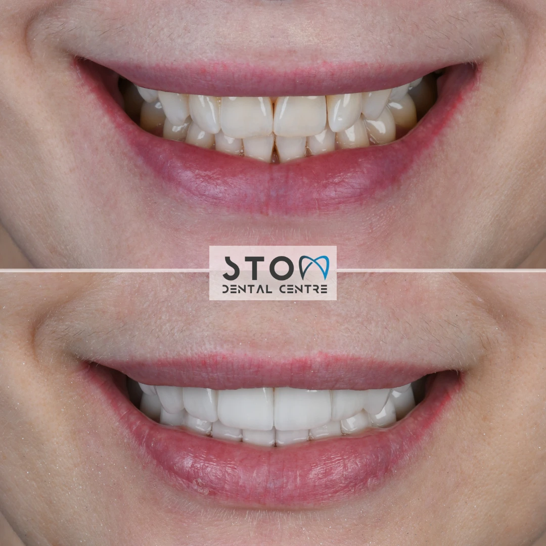
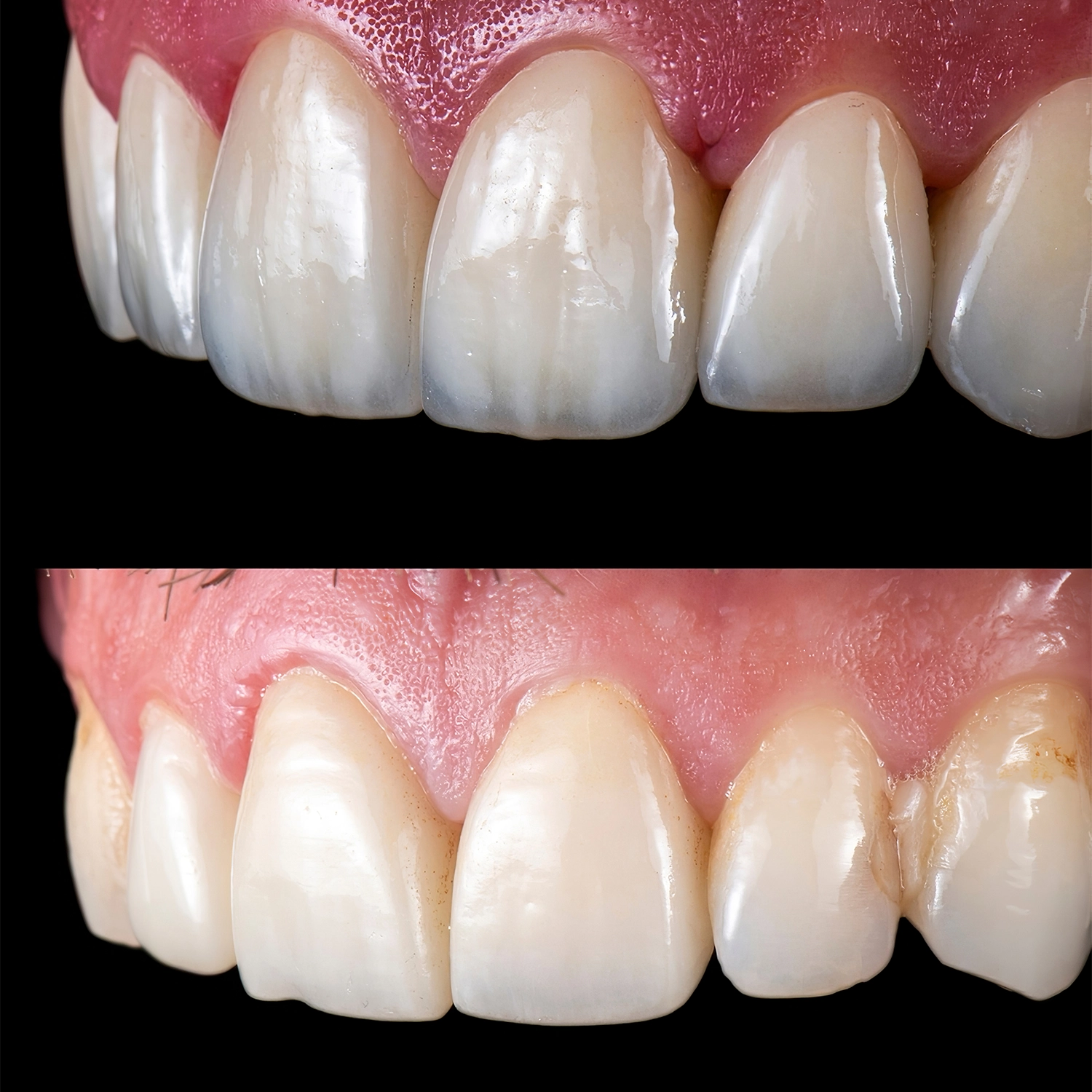
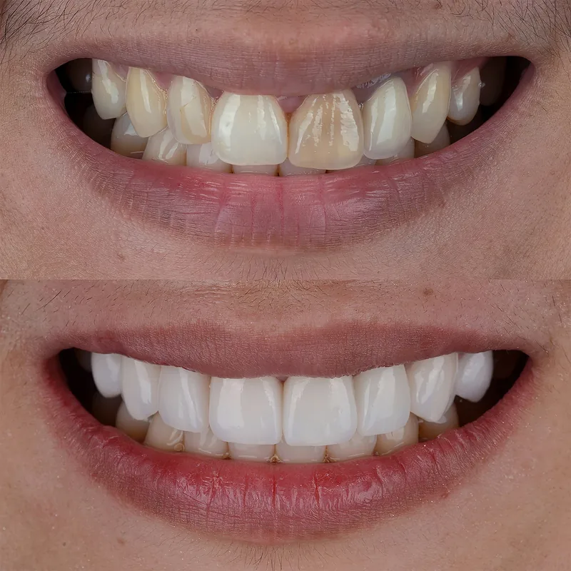
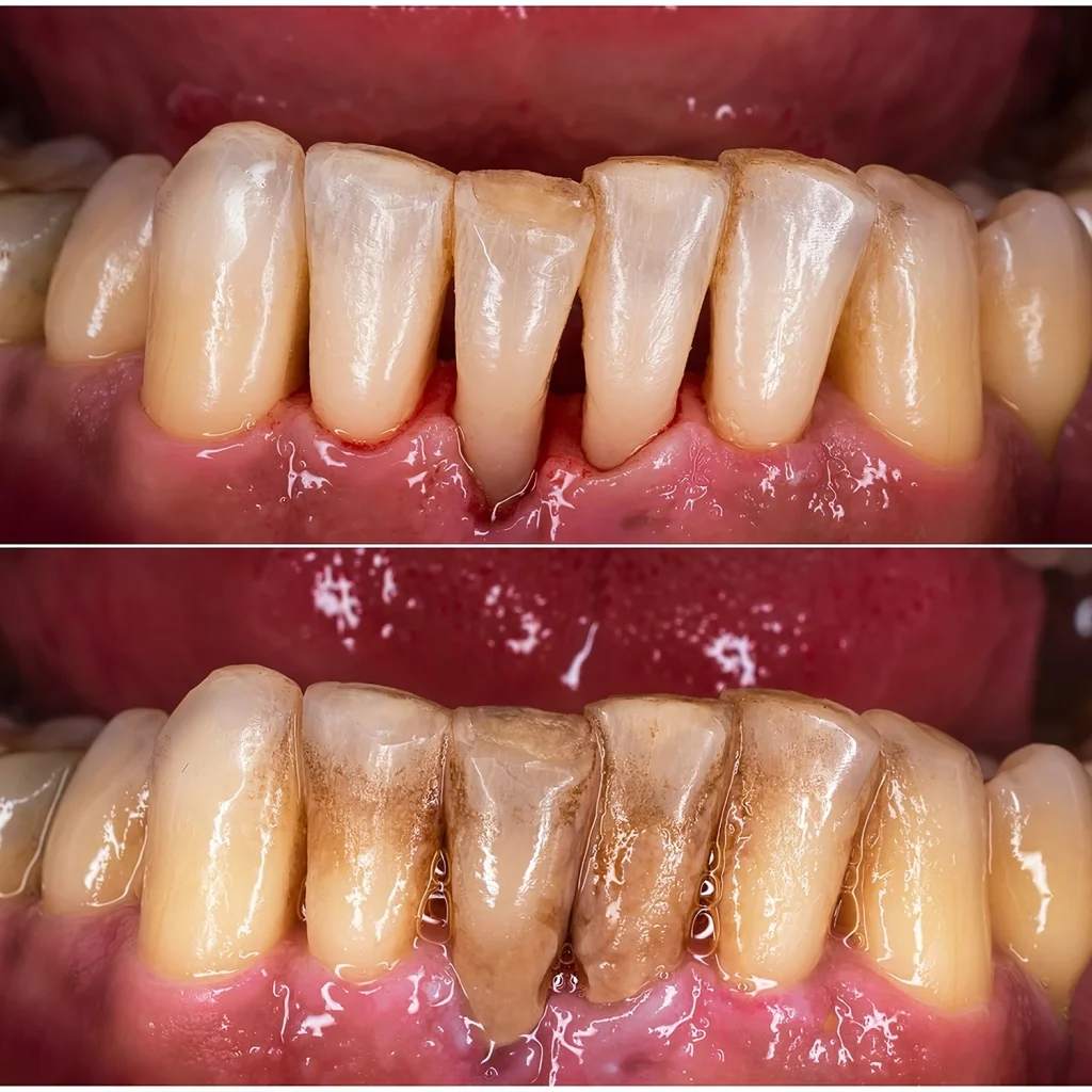
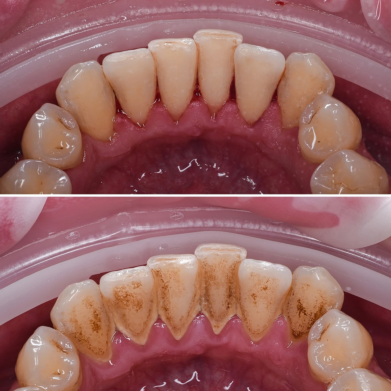
Before & after
Before and after: patients of our clinic

 BeforeAfter

 BeforeAfter

 BeforeAfter
Photos taken at our clinic: cropped only, not retouched. Every result is individual.
See all before and after cases