What Is a Dental Bridge? Types, Procedure and How Long One Lasts
Meet the Stom Dental Centre team
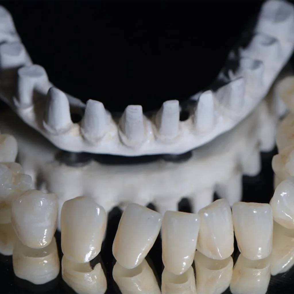
A dental bridge is a false tooth that is fixed to the teeth next to a gap. You cannot take it out yourself. Dentists call it a fixed partial denture; the official glossary of the specialty lists “bridge” as slang. It is one of the usual ways to replace one or two missing teeth.
What the short definitions leave out is the trade. A conventional bridge borrows strength from the neighbouring teeth, and those teeth pay for it: they are ground down, crowned, and carry a share of the risk for as long as the bridge is in your mouth. This article explains the parts and the types, what happens at each visit, what the long-term studies found, and where the evidence is thin. It does not tell you whether a bridge is right for you. Only an examination of your teeth, gums and bite can do that.
- What it is. An artificial tooth (the pontic) joined to crowns or wings on the neighbouring teeth (the abutments). You cannot take it out yourself.
- Four main types. Conventional, cantilever, resin-bonded (Maryland) and implant-supported. They differ in how much tooth is cut and in how they fail.
- How long it lasts depends on where you look. Pooled studies: 93.8% still in place at 5 years and 89.2% at 10. One specialist practice: 85% at 15 years. A university student clinic: 66.2% at 20 years.
- The supporting teeth carry the cost. In laboratory models a full-crown preparation removed about 63–76% of the crown of a tooth, and published rates of later nerve death range from about 2% to about one in three.
- Metal-ceramic has the best randomised evidence. In a meta-analysis of seven randomised trials, porcelain-veneered zirconia bridges failed more often than metal-ceramic ones. One manufacturer limits glass-ceramic bridges to three units, no further back than the second premolar.
- Cleaning under the false tooth is not optional. In an experiment from the 1980s, the gum under the pontic stayed healthy only when it was cleaned with floss every day.
The Parts of a Bridge, in Plain Words
Dentists use a handful of words when they plan a bridge, and it helps to know them before the consultation. The first four definitions below follow the Glossary of Prosthodontic Terms (10th edition, 2023).
| Term | What it means |
|---|---|
| Pontic | The artificial tooth that fills the gap and restores the function of the missing tooth |
| Abutment | A tooth, or an implant, that supports the bridge |
| Retainer | The part that joins the bridge to the abutment; in a conventional bridge, the crown on the supporting tooth |
| Connector | The joint between retainer and pontic; rigid, or built to allow limited movement |
| Unit | Each tooth position in the bridge; in the studies, a three-unit bridge replaces one tooth (our reading: two retainers and one pontic) |
The underside of the pontic matters more than it looks. The glossary describes several shapes. A hygienic pontic is convex near the gum and does not overlap the ridge. A modified ridge lap touches the ridge only on the visible side. An ovate pontic has an egg-shaped base that sits in a shallow dip in the gum, so the tooth seems to grow out of it. The old saddle shape, which wraps over both sides of the ridge, is the hardest to clean because food collects under it, and the textbooks summarised in one survey paper advise against it (Kazmi 2018). That paper reports textbook rules, not trial results: ovate or modified ridge lap for upper front teeth, hygienic shapes for lower back teeth.
Does a pontic that sits in close contact with the gum cause trouble? In a study of 12 patients with ovate pontics in the upper back teeth, fitted with tight contact but without pressure and cleaned underneath with floss once a day, only three sites showed mild inflammation after 12 months, although under the microscope the tissue was thinner than at control sites (Zitzmann 2002). Twelve patients for one year is a small base.
The Four Main Types, and a Fifth You May Hear About
The types differ in what holds the false tooth. The figures in the table come from different groups of patients and different decades. They are not a head-to-head comparison.
| Type | How it is held | Still in place |
|---|---|---|
| Conventional bridge | Crowns on the teeth at both ends of the gap | 93.8% at 5 years, 89.2% at 10 (Pjetursson 2007) |
| Cantilever bridge | Crowns on one side only; the false tooth hangs free | 91.4% at 5 years, 80.3% at 10 (Pjetursson 2007) |
| Resin-bonded (Maryland) bridge | A metal or ceramic wing glued to the back of a neighbouring tooth | 91.4% at 5 years, 82.9% at 10 (Thoma 2017) |
| Implant-supported bridge | Implants in the jaw; the neighbouring teeth are not touched | 95.4% at 5 years, 80.1% at 10; metal-ceramic only: 93.9% at 10 (Pjetursson 2012) |
| Inlay-retained bridge | Inlays in the neighbouring teeth instead of full crowns | 87.9% at 5 years (Chen 2017) |
Conventional bridge
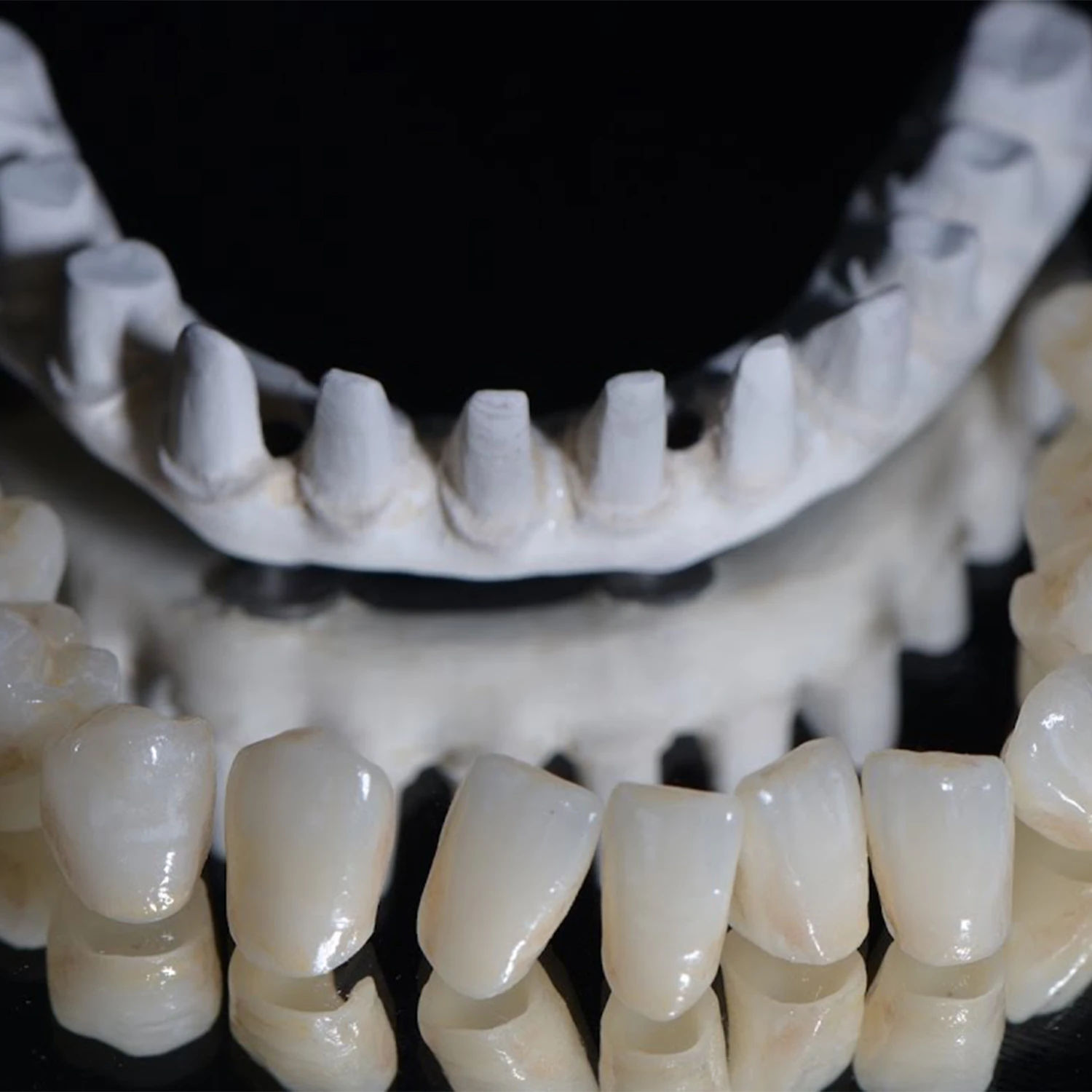
The teeth on both sides of the gap are shaped, each gets a crown, and the false tooth is joined to the two crowns. A leaflet from Bristol Dental School, which is patient information and not a study, says conventional bridges can replace only a small number of teeth, need strong neighbours, and are not used on teeth with active decay or gum disease.
Cantilever bridge
Here the false tooth is supported at one end only. A narrative review names the lever effect on the supporting teeth as the main drawback and says cantilevers do better at the front of the mouth, where biting forces are lower; the classic case is a small upper side tooth carried by the canine next to it (Sharma 2012a). In a study that re-examined 115 cantilever bridges 5 to 16 years after fitting, 10% of the supporting teeth that had been alive had lost their nerve, and a supporting tooth had fractured in 3%, twice as often at the tooth right next to the hanging part (Hämmerle 2000).
Resin-bonded (Maryland) bridge
A wing is bonded to the enamel of the neighbouring tooth, with little or no drilling. The Bristol leaflet for this type says it is usually for a single missing tooth, that the supporting tooth must be healthy, and that treatment usually takes two to three appointments of one to two hours. Its weak point is coming unstuck: about 15% debonded within five years in pooled data (Thoma 2017). The leaflet’s advice is practical: keep the bridge, because it can possibly be re-cemented. For back teeth the record is weaker. In one Hong Kong series of 101 resin-bonded bridges at the back of the mouth, 62.4% survived over a mean of about ten and a half years and a third had debonded; bridges fitted after 2001 did better, with 74.1% at ten years (Lam 2019).
Implant-supported bridge
When two or more teeth in a row are missing, implants can carry the bridge and the natural neighbours are left alone. Survival of these bridges was 95.4% at five years, but only 66.4% of patients had no complication of any kind in that time; chipped porcelain was the most frequent problem (Pjetursson 2012). Our guide to dental implants covers the surgery.
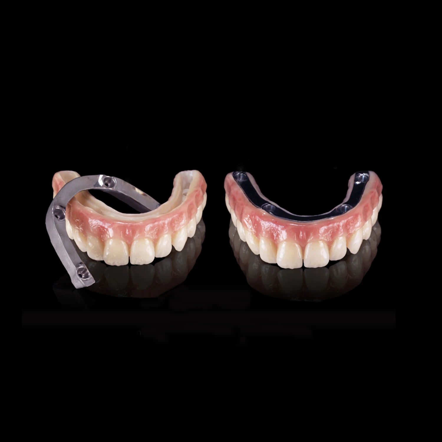
A bridge for a whole jaw is a different treatment with its own evidence. See our pages on the Toronto bridge and titanium-bar zirconia bridges and our comparison of full-arch options.
Inlay-retained bridge
This design holds the false tooth with inlays to save tooth substance. A systematic review estimated 87.9% survival at five years with more debonding and chipping than conventional bridges, and its authors call it a short- or medium-term option (Chen 2017). One German series of glass-ceramic inlay bridges did much worse: 22% were still in place after 15 years, and the authors could not recommend that design for regular use (Becker 2019).
How Many Teeth Can One Bridge Replace?
The traditional answer is Ante’s law. The glossary defines it as the observation that the root surface of the supporting teeth should be at least equal to that of the teeth being replaced, and credits it to a 1928 paper by the Toronto dentist Irwin Ante. It is a planning rule, not a tested limit. When researchers looked at bridges that deliberately broke it, on teeth with much reduced but healthy and well-maintained support, 96.4% of 579 bridges were in place at five years and 92.9% at ten (Lulic 2007). The review covers teeth whose support was much reduced but healthy; it does not cover untreated gum disease.
Length still counts in other data. In a Belgian university student clinic, 70.8% of short bridges survived 20 years against 52.8% of long ones; short bridges mostly failed for biological reasons such as decay, long ones for technical reasons (De Backer 2008). Among the same clinic’s bridges, three-unit ones reached 73.1% at 20 years and those with more than three units 61.5% (De Backer 2006b). A specialist practice with 515 metal-ceramic bridges found the opposite: the number of units made no difference (Walton 2002). And in a series of 30 long zirconia bridges of four to seven units, 16 had a failure or complication within five years and four had to be replaced (Schmitter 2012).
Our reading: there is no fixed number. Longer bridges ask more of the supporting teeth and of the material, and how much more depends on who makes them and from what.
Materials: Metal-Ceramic, Zirconia, Glass-Ceramic
| Material | What it is | What bridge studies found |
|---|---|---|
| Metal-ceramic | A metal frame covered with porcelain | 100% survival at 10 years against 91.3% for zirconia-ceramic in a randomised trial of 76 back-tooth bridges, 53 of them examined at 10 years; the authors rate most outcomes as similar (Sailer 2018) |
| Veneered zirconia | A zirconia frame covered with porcelain | More failures (risk ratio 3.64) and more chipping (2.92) than metal-ceramic across seven randomised trials (Chen 2024) |
| Monolithic zirconia | Zirconia through the full thickness, no porcelain layer | 87% at 5 years in one trial, against 97% for veneered zirconia and 100% for metal-ceramic (Pontevedra 2024) |
| Lithium disilicate (glass-ceramic) | A glass-ceramic of medium strength | 87.9% at 10 years, 48.6% at 15 years in the same group of bridges (Kern 2012; Garling 2019) |
Metal-ceramic is the reference material, and the pooled randomised trials against veneered zirconia still favour it (180 zirconia and 206 metal-ceramic bridges in seven trials). The authors of the meta-analysis conclude that the evidence supports a preference for metal-ceramic over veneered zirconia and that data beyond ten years are lacking for both kinds of zirconia bridge (Chen 2024). An early generation of veneered zirconia bridges at the back of the mouth did badly: estimated survival at 13 years was 43.2% for bridges supported at both ends and 52.5% for cantilevers, with wide margins of error, and of the 58 bridges 15 were lost to problems of the supporting teeth and 6 to failure of the ceramic (Passia 2019).
Monolithic zirconia has no porcelain layer that could chip. In a review of 6,370 zirconia restorations, minor chipping at five years was estimated at 10.4% for veneered and 0.08% for monolithic zirconia (Shihabi 2023). But most restorations in that review were single crowns, not bridges, and in one randomised trial of three-unit bridges (30 per group) the monolithic group had the lowest survival of the three (87% against 97% and 100%), although its authors still call monolithic zirconia a possible alternative; most complications in that trial were biological (Pontevedra 2024). The data on monolithic zirconia bridges are still few.
Lithium disilicate is the cautionary case. The same group of 36 three-unit bridges that looked comparable to metal-ceramic at ten years had 48.6% survival at fifteen; the authors put the late failures down to fatigue of the ceramic (Garling 2019). The instructions for use of one widely used product (IPS e.max CAD, edition of April 2022) allow three-unit bridges only up to the second premolar as the rearmost support, and list cantilever, inlay and Maryland bridges and untreated teeth grinding under limitations of use. That is a manufacturer’s document for one product, not independent evidence.
A Cochrane review of nine trials, all rated as very low-quality evidence, found too little to say that metal-free materials are better or worse than metal-ceramic, and advised dentists to keep choosing by their clinical experience and the patient’s circumstances and preferences (Poggio 2017). Our pages on zirconia crowns and e.max against zirconia go into the materials for single teeth.
The Procedure, Visit by Visit
The steps below come from patient leaflets of UK dental hospitals and the Australian government health portal healthdirect. They describe a typical course; they are not study results, and your own plan may differ.
- Assessment. The mouth is examined, X-rays check the supporting teeth, and decay or gum disease is treated first (healthdirect).
- Preparation. Under local anaesthetic the teeth next to the gap are shaped. One hospital leaflet gives one to two hours in the chair for this visit (University Hospitals of North Midlands).
- Impression and colour. A mould goes to the dental laboratory, and the shade is chosen. Some practices use a digital scan in place of the mould (see below).
- Temporary bridge. It protects the prepared teeth until the final bridge is ready.
- Fitting. The temporary bridge comes off, the final one is tried in and cemented, and the bite is adjusted (Bristol leaflet). The North Midlands leaflet puts this visit two to three weeks after the first and at up to an hour.
- Review. A check-up follows, and the hospital arranges a hygiene appointment to show how to clean the bridge.
The Bristol leaflet counts two to three visits. In a Swiss practice comparison, a three-unit bridge took 4.8 visits on average and an implant crown 8.1, counting preparatory treatment and later complication visits, with similar total chair time (Brägger 2005). If you travel for treatment, ask for the visit plan in writing.
Scan or impression? In a review of 17 studies, scanning was faster and patients generally preferred it, with similar results for the finished work (Siqueira 2021). A summary of eleven reviews found scans as accurate as impressions for fixed work of fewer than four units and recommended conventional impressions for more extensive work (Afrashtehfar 2022).

Two details are worth knowing. First, the nerve of a prepared tooth can die without symptoms before the crown or bridge is even cemented. In a study of 120 teeth prepared for crowns this happened in 9%: in 5% of teeth that had been intact and 13% of teeth that already had decay, fillings or crowns (Kontakiotis 2015). The teeth were tested before cementation and treated first where needed. Second, the cement may matter. In one 15-year recall, crowns and bridge retainers fixed with an adhesive resin survived 90.5% against 67.4% with conventional cements, with far less decay underneath (Piemjai 2022). That was one centre and not a randomised comparison.
What will it feel like afterwards? The Bristol leaflet says the tooth may be sensitive at first, that the new bridge feels unusual, and that patients quickly get used to it. In a trial of 120 crowned teeth with living nerves, 6–13% were sensitive one week after cementation, depending on the cement, and none at two years (Denner 2007).
Can a bridge go in straight after an extraction? We found no source that gives a waiting time for the final bridge. What is documented is that the ridge shrinks: on average 3.79 mm of width within six months (Tan 2012). A review article adds that the healed ridge may become concave, so that a false tooth looks as if it sits on the gum instead of growing out of it (Brennand Roper 2024). Temporary replacements are used in the meantime.
What a Bridge Asks of the Supporting Teeth
The preparation cannot be undone. In laboratory models, shaping a tooth for a full crown removed about 63–72% of the crown of a front tooth and 68–76% of a back tooth; veneer and resin-bonded wing preparations on front teeth needed about 3–30% (Edelhoff 2002).
How often does the nerve of a supporting tooth die later, so that it needs a root canal treatment? This is the question where the sources disagree most.
| Source | Setting | Finding |
|---|---|---|
| Cheung 2005 | Hong Kong teaching hospital, about 15 years | Nerve death in 32.5% of bridge-supporting teeth, against 15.6% of teeth with single crowns |
| Valderhaug 1997 | Oslo, crowns and bridges made by senior students; 28% of the patients still in the study at 25 years | 92% of living crowned teeth still free of nerve damage at 10 years, 83% at 25 |
| Hämmerle 2000 | Cantilever bridges, 5 to 16 years | 10% of living supporting teeth lost their nerve |
| Walton 2002 | One specialist practice, up to 15 years | 2% of living supporting teeth later needed a root canal treatment |
The patient leaflets vary just as much: “roughly 20%” in Bristol, “a small percentage” in Manchester. A fair summary is that the risk is real, grows with time, was higher for bridge supports than for single crowns in the one direct comparison, and has been measured at anything from a few per cent to about one in three. Our article on root canal treatment and crowns explains what the treatment involves.
Decay is the other long-term threat. It was the main reason for failure of crowned teeth in the Oslo group, in 12% of the teeth over 25 years (Valderhaug 1997). In the well-maintained periodontal patients mentioned above, 98.1% of supporting teeth were free of decay at ten years (Lulic 2007). The edge of the crown counts as well: in a ten-year follow-up, gums were more inflamed where the crown edge lay below the gum line than where it lay at or above it (Valderhaug 1980).
How Long Bridges Last, and Why They Fail
| Where the bridges were made | Result |
|---|---|
| Pooled cohort studies | 93.8% in place at 5 years, 89.2% at 10 (Pjetursson 2007); but only 71.1% free of any complication at 10 (Tan 2004) |
| One specialist practice, 515 metal-ceramic bridges | 96% at 5 years, 87% at 10, 85% at 15 (Walton 2002) |
| University student clinic, Belgium, 322 bridges | 66.2% at 20 years (De Backer 2006a) |
| University student clinic, Finland | 78% of the 82 bridges examined after a mean of 17.7 years; 46 of the original 128 were not examined (Näpänkangas 2011) |
| NHS general dental services, England and Wales | 72% of the teeth supporting conventional bridges went 10 years without further treatment other than re-cementing (Burke 2012) |
A UK hospital leaflet tells patients that crowns and bridges last “five to ten years” on average, with some lasting twenty or thirty. The cohort data are better than that, and the honest answer is a range. Our reading: the setting, the material and the upkeep explain more of the differences than the word “bridge” does.
Why do they fail? Decay was the leading cause in the Belgian student clinic and in a small American study, but not in the specialist practice. In the Belgian clinic, decay and bridges coming loose made up 61% of failures (De Backer 2006a). In the American recall study decay caused 5 of 13 failures (38%), and those bridges had served 16 years on average before it did (Libby 1997). In the specialist practice the order was different. Of the bridges that needed retreatment, tooth fracture accounted for 38%, gum and bone breakdown for 27%, loss of retention for 13% and decay for 11% (Walton 2003). Root-treated teeth were weaker supports in some series but not in all: in the lower jaw, three-unit bridges on living teeth reached 96.3% at 20 years against 69.3% on root-treated teeth, while bridges on living teeth in the upper jaw reached 70.2% (De Backer 2006b); the Oslo study found no difference (Valderhaug 1997). In the specialist practice, regular professional maintenance went with significantly fewer failures (Walton 2003). That is an association; people who keep appointments may differ in other ways.
What can be repaired? A bridge that has come loose but is otherwise sound may be re-cemented, once the dentist has found out why it came loose (Sharma 2012b). Chipped porcelain is less clear. The Bristol leaflet says a fractured bridge will need to be replaced; a literature review describes polishing or rebuilding the chip with composite in the mouth as a temporary but effective solution; 17 of its 21 studies were laboratory studies, so the clinical data are few (Alqarawi 2022). Taking off a firmly cemented bridge is often destructive (Sharma 2012b).
How does a bridge compare with an implant? In one specialist practice over 15 years, the survival of metal-ceramic three-unit bridges and implant crowns did not differ significantly at the back of the mouth (92.75% against 95.95%), while at the front the implant crowns did better (93.33% against 82.82%). Seventeen bridges that the dentist judged to have a poor outlook at fitting were left out of the comparison (Walton 2015). That is one specialist practice, not a randomised trial. Our article implant, bridge or denture for a missing tooth sets the options side by side.
Planning dental treatment in Antalya?
Get a free, no-obligation assessment from our clinical team — we reply within 24 hours.
Cleaning Under a Bridge
The crowns of a bridge are joined, so floss cannot be passed between them in the normal way (healthdirect). The space under the false tooth needs its own cleaning.
The direct evidence is old. In a Danish experiment from the 1980s, the gum under the pontic became inflamed when the area was not cleaned, or was cleaned with a toothbrush and toothpicks only, and stayed healthy when it was cleaned with floss every day (Tolboe 1987). A follow-up with five patients found that the material of the pontic mattered less than the cleaning (Tolboe 1988). We found no trial that compares floss threaders, superfloss, interdental brushes and water flossers under a bridge.
So the advice rests on general evidence and on what dental teams teach:
- Brush the bridge like your own teeth, twice a day with fluoride toothpaste (healthdirect; Bristol Dental School leaflet).
- Clean underneath every day with the aid your dental team shows you. healthdirect names superfloss, floss threaders and interdental brushes.
- A Cochrane review of 35 trials found that floss or interdental brushes in addition to brushing may reduce gum inflammation or plaque more than brushing alone, and that interdental brushes may work better than floss; the certainty was low to very low and the effects were small (Worthington 2019).
- The European periodontal guideline recommends interdental brushes where they fit, for patients in periodontal aftercare (Sanz 2020).
- For water flossers we found studies around implants only, none on tooth-supported bridges.
Bad breath has been studied little. One study of 48 bridge wearers linked it to designs that are hard to clean, such as saddle-shaped pontics (Zigurs 2005).
How often should you be checked? We found no professional body that sets an interval for people with bridges. The NICE guideline for England and Wales sets the recall individually, between 3 and 24 months for adults, according to disease and risk (NICE 2004). If plaque and hard deposits are your problem, our guide to dental calculus explains how they form.
When a Bridge Is Not the Best Idea
The points below come from the risk patterns in the studies above. They are not a guideline checklist.
- Untouched neighbours. The Bristol leaflet says conventional bridges are not used on teeth that have never had a filling. Our reading: having two healthy teeth cut to replace one is the main reason to ask about an implant or a resin-bonded bridge first.
- Weak or root-treated supports. They failed more often in several series (De Backer 2006b; Walton 2002).
- Active decay or untreated gum disease. Both are treated first. Bridges on teeth with reduced support did well only in treated and maintained patients (Lulic 2007); in patients with stage IV periodontitis who had been treated first, a review of mostly case series found that 4.8% of supporting teeth were lost (Montero 2022). Our guide to the stages of periodontitis explains what a diagnosis means.
- Long gaps. Long bridges did worse in the Belgian student clinic, and length was a risk factor in a series of long zirconia bridges, while one specialist practice found no effect of the number of units (see above). The manufacturer’s instructions quoted above do not cover long spans for glass-ceramic.
- Front teeth as supports. Upper front teeth had the highest rate of nerve death in the Hong Kong study of 77 supporting teeth (Cheung 2005), and front supports went with more failures in the specialist practice (Walton 2002).
- Teeth grinding. The evidence is mixed. A systematic review of ceramic restorations in general found more failures of front-tooth veneers in people with sleep bruxism but no overall link for other ceramic restorations (odds ratio 1.10), with the quality of evidence rated very low (de Souza Melo 2018), while a manufacturer lists untreated bruxism as a limitation. See our article on teeth grinding and dental work.
- Which type do you recommend for my gap, and why not the others?
- How much of each supporting tooth will be removed, and what condition are those teeth in today?
- What material will the bridge be made of, and what is its record for a bridge of this length in this part of the mouth?
- How will you check the nerves of the supporting teeth before the bridge is cemented?
- What shape will the false tooth have underneath, and how do I clean it?
- How many visits are needed, and what happens if the bridge comes loose or chips?
Frequently Asked Questions
Is getting a dental bridge painful?
The teeth are prepared under local anaesthetic. We found no study that measured pain during bridge treatment. Afterwards some teeth are sensitive for a while: in one trial 6–13% of crowned teeth were sensitive after a week, depending on the cement, and none after two years; two of the 120 teeth needed a root canal treatment (Denner 2007).
How long does a dental bridge last?
In pooled studies 93.8% were in place at 5 years and 89.2% at 10. One specialist practice reported 85% at 15 years, a student clinic 66.2% at 20. The range reflects different settings, materials and upkeep.
Does a bridge damage the neighbouring teeth?
A conventional bridge requires grinding them down, which cannot be undone: about 63–76% of the crown in laboratory models. Some supporting teeth later need a root canal treatment. Published figures range from about 2% to about one in three, depending on the study and the follow-up.
Can a bridge be put on front teeth?
Yes. Conventional, cantilever and resin-bonded bridges are all used at the front. Two cautions from the studies: in one study upper front teeth used as supports lost their nerve more often, and in one practice implant crowns outlasted three-unit bridges at the front over 15 years.
Can a dental bridge be whitened?
No. Whitening only lightens natural teeth (NHS.uk, which does not recommend it for people with crowns, dentures or implants), and a UK hospital leaflet notes that the porcelain on crowns is not affected by bleaching. The same applies to the porcelain of a bridge, which will then no longer match whitened natural teeth.
Can a bridge be removed or re-cemented?
A bridge is fixed, and you cannot take it out yourself. A loose but sound bridge may be re-cemented once the cause is known. Removing a firmly cemented bridge is often destructive, so a new one may be needed afterwards (Sharma 2012b).
How do I clean under a bridge?
Every day, with the aid your dental team shows you: superfloss, a floss threader or an interdental brush. In an experiment from the 1980s the gum under the false tooth stayed healthy only with daily flossing (Tolboe 1987). No trial has compared the modern aids under a bridge.
Is zirconia better than metal-ceramic for a bridge?
Not on current evidence. Across seven randomised trials, porcelain-veneered zirconia bridges failed and chipped more often than metal-ceramic ones (Chen 2024). Monolithic zirconia chips far less, but bridge-specific data are few and one trial was unfavourable.
Sources
- Layton DM, Morgano SM, Att W, et al. (eds). The Glossary of Prosthodontic Terms 2023: Tenth Edition. J Prosthet Dent. 2023;130(4 Suppl 1):e7-e126. doi:10.1016/j.prosdent.2023.03.002. PMID 37914442.
- Kazmi SMR, Iqbal Z, Muneer MU, Riaz S, Zafar MS. Different pontic design for porcelain fused to metal fixed dental prosthesis: contemporary guidelines and practice by general dental practitioners. Eur J Dent. 2018;12(3):375-379. doi:10.4103/ejd.ejd_232_18. PMID 30147402.
- Zitzmann NU, Marinello CP, Berglundh T. The ovate pontic design: a histologic observation in humans. J Prosthet Dent. 2002;88(4):375-380. doi:10.1067/mpr.2002.128758. PMID 12447213.
- Pjetursson BE, Brägger U, Lang NP, Zwahlen M. Comparison of survival and complication rates of tooth-supported fixed dental prostheses (FDPs) and implant-supported FDPs and single crowns (SCs). Clin Oral Implants Res. 2007;18 Suppl 3:97-113. doi:10.1111/j.1600-0501.2007.01439.x. PMID 17594374.
- Thoma DS, Sailer I, Ioannidis A, Zwahlen M, Makarov N, Pjetursson BE. A systematic review of the survival and complication rates of resin-bonded fixed dental prostheses after a mean observation period of at least 5 years. Clin Oral Implants Res. 2017;28(11):1421-1432. doi:10.1111/clr.13007. PMID 28191679.
- Pjetursson BE, Thoma D, Jung R, Zwahlen M, Zembic A. A systematic review of the survival and complication rates of implant-supported fixed dental prostheses (FDPs) after a mean observation period of at least 5 years. Clin Oral Implants Res. 2012;23 Suppl 6:22-38. doi:10.1111/j.1600-0501.2012.02546.x. PMID 23062125.
- Chen J, Cai H, Suo L, Xue Y, Wang J, Wan Q. A systematic review of the survival and complication rates of inlay-retained fixed dental prostheses. J Dent. 2017;59:2-10. doi:10.1016/j.jdent.2017.02.006. PMID 28212978.
- Becker M, Chaar MS, Garling A, Kern M. Fifteen-year outcome of posterior all-ceramic inlay-retained fixed dental prostheses. J Dent. 2019;89:103174. doi:10.1016/j.jdent.2019.07.012. PMID 31362035.
- University of Bristol, Bristol Dental School. Conventional dental bridges and Resin-bonded bridges: patient information leaflets. bristol.ac.uk, accessed 21 November 2026.
- Sharma A, Rahul GR, Poduval ST, Shetty K. Assessment of various factors for feasibility of fixed cantilever bridge: a review study. ISRN Dent. 2012;2012:259891. doi:10.5402/2012/259891. PMID 22461987. (Sharma 2012a) And by the same authors: Removal of failed crown and bridge. J Clin Exp Dent. 2012;4(3):e167-e172. doi:10.4317/jced.50690. PMID 24558549. (Sharma 2012b)
- Hämmerle CH, Ungerer MC, Fantoni PC, Brägger U, Bürgin W, Lang NP. Long-term analysis of biologic and technical aspects of fixed partial dentures with cantilevers. Int J Prosthodont. 2000;13(5):409-415. PMID 11203663.
- Lam WYH, Chan RST, Li KY, Tang KT, Lui TT, Botelho MG. Ten-year clinical evaluation of posterior fixed-movable resin-bonded fixed partial dentures. J Dent. 2019;86:118-125. doi:10.1016/j.jdent.2019.06.003. PMID 31181243.
- Lulic M, Brägger U, Lang NP, Zwahlen M, Salvi GE. Ante’s (1926) law revisited: a systematic review on survival rates and complications of fixed dental prostheses (FDPs) on severely reduced periodontal tissue support. Clin Oral Implants Res. 2007;18 Suppl 3:63-72. doi:10.1111/j.1600-0501.2007.01438.x. PMID 17594371.
- De Backer H, Van Maele G, De Moor N, Van den Berghe L, De Boever J. A 20-year retrospective survival study of fixed partial dentures. Int J Prosthodont. 2006;19(2):143-153. PMID 16602362. (De Backer 2006a)
- De Backer H, Van Maele G, De Moor N, Van den Berghe L. Single-tooth replacement: is a 3-unit fixed partial denture still an option? A 20-year retrospective study. Int J Prosthodont. 2006;19(6):567-573. PMID 17165295. (De Backer 2006b)
- De Backer H, Van Maele G, De Moor N, Van den Berghe L. Long-term results of short-span versus long-span fixed dental prostheses: an up to 20-year retrospective study. Int J Prosthodont. 2008;21(1):75-85. PMID 18350953.
- Walton TR. An up to 15-year longitudinal study of 515 metal-ceramic FPDs: Part 1. Outcome. Int J Prosthodont. 2002;15(5):439-445. PMID 12375457. Part 2. Modes of failure and influence of various clinical characteristics. Int J Prosthodont. 2003;16(2):177-182. PMID 12737251.
- Walton TR. An up-to-15-year comparison of the survival and complication burden of three-unit tooth-supported fixed dental prostheses and implant-supported single crowns. Int J Oral Maxillofac Implants. 2015;30(4):851-861. doi:10.11607/jomi.4220. PMID 26252025.
- Schmitter M, Mussotter K, Rammelsberg P, Gabbert O, Ohlmann B. Clinical performance of long-span zirconia frameworks for fixed dental prostheses: 5-year results. J Oral Rehabil. 2012;39(7):552-557. doi:10.1111/j.1365-2842.2012.02311.x. PMID 22574689.
- Sailer I, Balmer M, Hüsler J, Hämmerle CHF, Känel S, Thoma DS. 10-year randomized trial (RCT) of zirconia-ceramic and metal-ceramic fixed dental prostheses. J Dent. 2018;76:32-39. doi:10.1016/j.jdent.2018.05.015. PMID 29807060.
- Chen H, Li T, Ng JPZ, Almeheni L, Li KY, Burrow MF. Clinical performance of zirconia-based tooth-supported fixed dental prostheses: a systematic review and meta-analysis. J Dent. 2024;151:105382. doi:10.1016/j.jdent.2024.105382. PMID 39369882.
- Pontevedra P, Lopez-Suarez C, Rodriguez V, Tobar C, Pelaez J, Suarez MJ. Digital workflow for monolithic and veneered zirconia and metal-ceramic posterior fixed partial dentures: a five-year prospective randomized clinical trial. J Prosthodont Res. 2024;68(1):78-84. doi:10.2186/jpr.JPR_D_22_00237. PMID 36990752.
- Passia N, Chaar MS, Kern M. Outcome of posterior fixed dental prostheses made from veneered zirconia over an observation period of up to 13 years. J Dent. 2019;86:126-129. doi:10.1016/j.jdent.2019.05.032. PMID 31150728.
- Shihabi S, Chrcanovic BR. Clinical outcomes of tooth-supported monolithic zirconia vs. porcelain-veneered zirconia fixed dental prosthesis, with an additional focus on the cement type: a systematic review and meta-analysis. Clin Oral Investig. 2023;27(10):5755-5769. doi:10.1007/s00784-023-05219-4. PMID 37626273.
- Kern M, Sasse M, Wolfart S. Ten-year outcome of three-unit fixed dental prostheses made from monolithic lithium disilicate ceramic. J Am Dent Assoc. 2012;143(3):234-240. doi:10.14219/jada.archive.2012.0147. PMID 22383203.
- Garling A, Sasse M, Becker MEE, Kern M. Fifteen-year outcome of three-unit fixed dental prostheses made from monolithic lithium disilicate ceramic. J Dent. 2019;89:103178. doi:10.1016/j.jdent.2019.08.001. PMID 31394121.
- Ivoclar Vivadent AG. IPS e.max CAD: instructions for use (revision 4, information prepared 11 April 2022). Manufacturer document, read from a distributor-hosted copy on 21 November 2026.
- Poggio CE, Ercoli C, Rispoli L, Maiorana C, Esposito M. Metal-free materials for fixed prosthodontic restorations. Cochrane Database Syst Rev. 2017;12(12):CD009606. doi:10.1002/14651858.CD009606.pub2. PMID 29261853.
- healthdirect Australia. Dental bridge. Last reviewed February 2025. healthdirect.gov.au, accessed 21 November 2026.
- University Hospitals of North Midlands NHS Trust, Restorative Department. Crowns and bridges: patient information leaflet (reapproved October 2025). uhnm.nhs.uk, accessed 21 November 2026.
- Manchester University NHS Foundation Trust, University Dental Hospital of Manchester. Crowns, veneers and bridges: information for patients (updated July 2018). mft.nhs.uk, accessed 21 November 2026.
- Brägger U, Krenander P, Lang NP. Economic aspects of single-tooth replacement. Clin Oral Implants Res. 2005;16(3):335-341. doi:10.1111/j.1600-0501.2005.01112.x. PMID 15877754.
- Siqueira R, Galli M, Chen Z, et al. Intraoral scanning reduces procedure time and improves patient comfort in fixed prosthodontics and implant dentistry: a systematic review. Clin Oral Investig. 2021;25(12):6517-6531. doi:10.1007/s00784-021-04157-3. PMID 34568955.
- Afrashtehfar KI, Alnakeb NA, Assery MKM. Accuracy of intraoral scanners versus traditional impressions: a rapid umbrella review. J Evid Based Dent Pract. 2022;22(3):101719. doi:10.1016/j.jebdp.2022.101719. PMID 36162879.
- Kontakiotis EG, Filippatos CG, Stefopoulos S, Tzanetakis GN. A prospective study of the incidence of asymptomatic pulp necrosis following crown preparation. Int Endod J. 2015;48(6):512-517. doi:10.1111/iej.12340. PMID 24964352.
- Piemjai M, Adunphichet N. Impact of hybrid layer formation on the 15-year survival, complications, and failures of full-coverage retainers. J Prosthodont Res. 2022;66(1):131-140. doi:10.2186/jpr.JPR_D_20_00249. PMID 34321373.
- Denner N, Heydecke G, Gerds T, Strub JR. Clinical comparison of postoperative sensitivity for an adhesive resin cement containing 4-META and a conventional glass-ionomer cement. Int J Prosthodont. 2007;20(1):73-78. PMID 17319368.
- Tan WL, Wong TL, Wong MC, Lang NP. A systematic review of post-extractional alveolar hard and soft tissue dimensional changes in humans. Clin Oral Implants Res. 2012;23 Suppl 5:1-21. doi:10.1111/j.1600-0501.2011.02375.x. PMID 22211303.
- Brennand Roper M, Fields Y. Achieving the optimal emergence profile: the role of soft tissue grafting and pontic site development. Br Dent J. 2024;237(11):843-849. doi:10.1038/s41415-024-8023-2. PMID 39672854.
- Edelhoff D, Sorensen JA. Tooth structure removal associated with various preparation designs for anterior teeth. J Prosthet Dent. 2002;87(5):503-509. doi:10.1067/mpr.2002.124094. PMID 12070513. And for posterior teeth: Int J Periodontics Restorative Dent. 2002;22(3):241-249. PMID 12186346.
- Cheung GS, Lai SC, Ng RP. Fate of vital pulps beneath a metal-ceramic crown or a bridge retainer. Int Endod J. 2005;38(8):521-530. doi:10.1111/j.1365-2591.2005.00982.x. PMID 16011770.
- Valderhaug J, Jokstad A, Ambjørnsen E, Norheim PW. Assessment of the periapical and clinical status of crowned teeth over 25 years. J Dent. 1997;25(2):97-105. doi:10.1016/s0300-5712(96)00008-5. PMID 9105139.
- Valderhaug J. Periodontal conditions and carious lesions following the insertion of fixed prostheses: a 10-year follow-up study. Int Dent J. 1980;30(4):296-304. PMID 6935162.
- Tan K, Pjetursson BE, Lang NP, Chan ES. A systematic review of the survival and complication rates of fixed partial dentures (FPDs) after an observation period of at least 5 years. Clin Oral Implants Res. 2004;15(6):654-666. doi:10.1111/j.1600-0501.2004.01119.x. PMID 15533126.
- Näpänkangas R, Raustia A. An 18-year retrospective analysis of treatment outcomes with metal-ceramic fixed partial dentures. Int J Prosthodont. 2011;24(4):314-319. PMID 21716968.
- Burke FJ, Lucarotti PS. Ten year survival of bridges placed in the General Dental Services in England and Wales. J Dent. 2012;40(11):886-895. doi:10.1016/j.jdent.2012.07.002. PMID 22864053.
- Libby G, Arcuri MR, LaVelle WE, Hebl L. Longevity of fixed partial dentures. J Prosthet Dent. 1997;78(2):127-131. doi:10.1016/s0022-3913(97)70115-x. PMID 9260128.
- Alqarawi FK, Alhumaidan AA, Aldakhili A, et al. Intraoral repair of fractured ceramics: a literature review. F1000Res. 2022;11:1275. doi:10.12688/f1000research.126547.1. PMID 39635089.
- Tolboe H, Isidor F, Budtz-Jörgensen E, Kaaber S. Influence of oral hygiene on the mucosal conditions beneath bridge pontics. Scand J Dent Res. 1987;95(6):475-482. doi:10.1111/j.1600-0722.1987.tb01963.x. PMID 3321391. And: Influence of pontic material on alveolar mucosal conditions. Scand J Dent Res. 1988;96(5):442-447. doi:10.1111/j.1600-0722.1988.tb01581.x. PMID 3059449.
- Worthington HV, MacDonald L, Poklepovic Pericic T, et al. Home use of interdental cleaning devices, in addition to toothbrushing, for preventing and controlling periodontal diseases and dental caries. Cochrane Database Syst Rev. 2019;4(4):CD012018. doi:10.1002/14651858.CD012018.pub2. PMID 30968949.
- Sanz M, Herrera D, Kebschull M, et al. Treatment of stage I-III periodontitis: the EFP S3 level clinical practice guideline. J Clin Periodontol. 2020;47 Suppl 22:4-60. doi:10.1111/jcpe.13290. PMID 32383274.
- Zigurs G, Vidzis A, Brinkmane A. Halitosis manifestation and prevention means for patients with fixed teeth dentures. Stomatologija. 2005;7(1):3-6. PMID 16254469.
- NHS.uk. Teeth whitening. Page last reviewed 19 August 2025. nhs.uk, accessed 21 November 2026.
- National Institute for Health and Care Excellence (NICE). Dental checks: intervals between oral health reviews. Clinical guideline CG19, 27 October 2004. nice.org.uk.
- Montero E, Molina A, Palombo D, Morón B, Pradíes G, Sanz-Sánchez I. Efficacy and risks of tooth-supported prostheses in the treatment of partially edentulous patients with stage IV periodontitis: a systematic review and meta-analysis. J Clin Periodontol. 2022;49 Suppl 24:182-207. doi:10.1111/jcpe.13482. PMID 34786742.
- de Souza Melo G, Batistella EÂ, Bertazzo-Silveira E, et al. Association of sleep bruxism with ceramic restoration failure: a systematic review and meta-analysis. J Prosthet Dent. 2018;119(3):354-362. doi:10.1016/j.prosdent.2017.07.005. PMID 28967401.
This article is for general information only and does not replace an individual dental examination or the advice of your treating dentist. The figures are estimates from different studies, countries and decades, many of them from specialist or university settings; they are not a guarantee of any outcome for any individual patient or clinic.
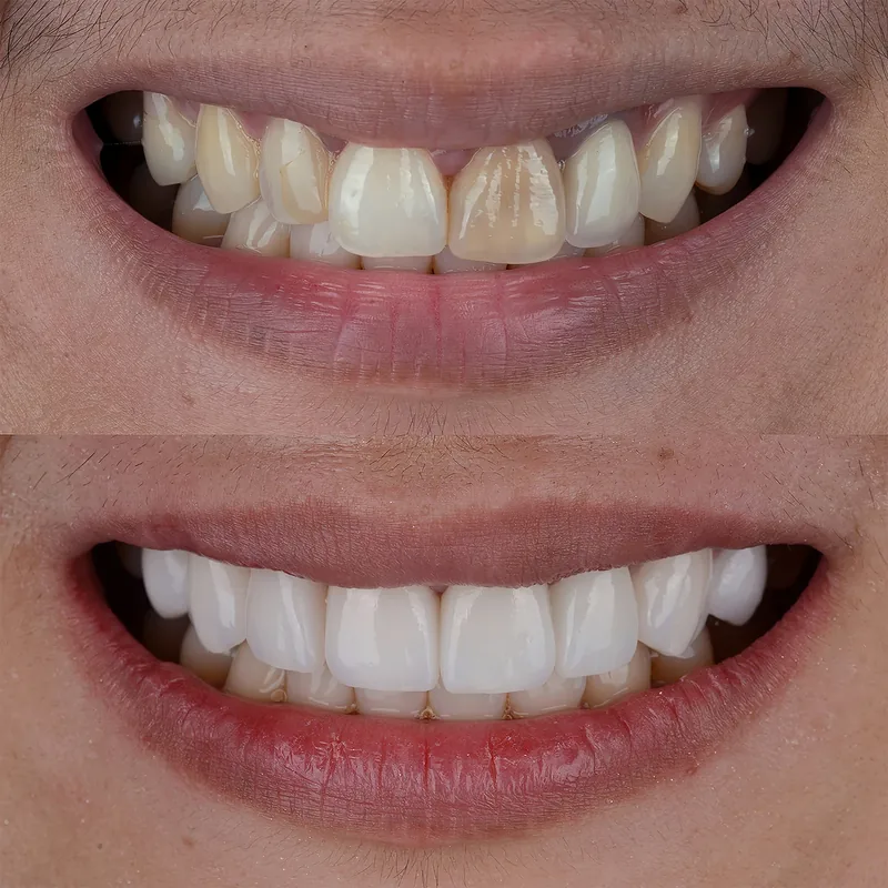
Before & after
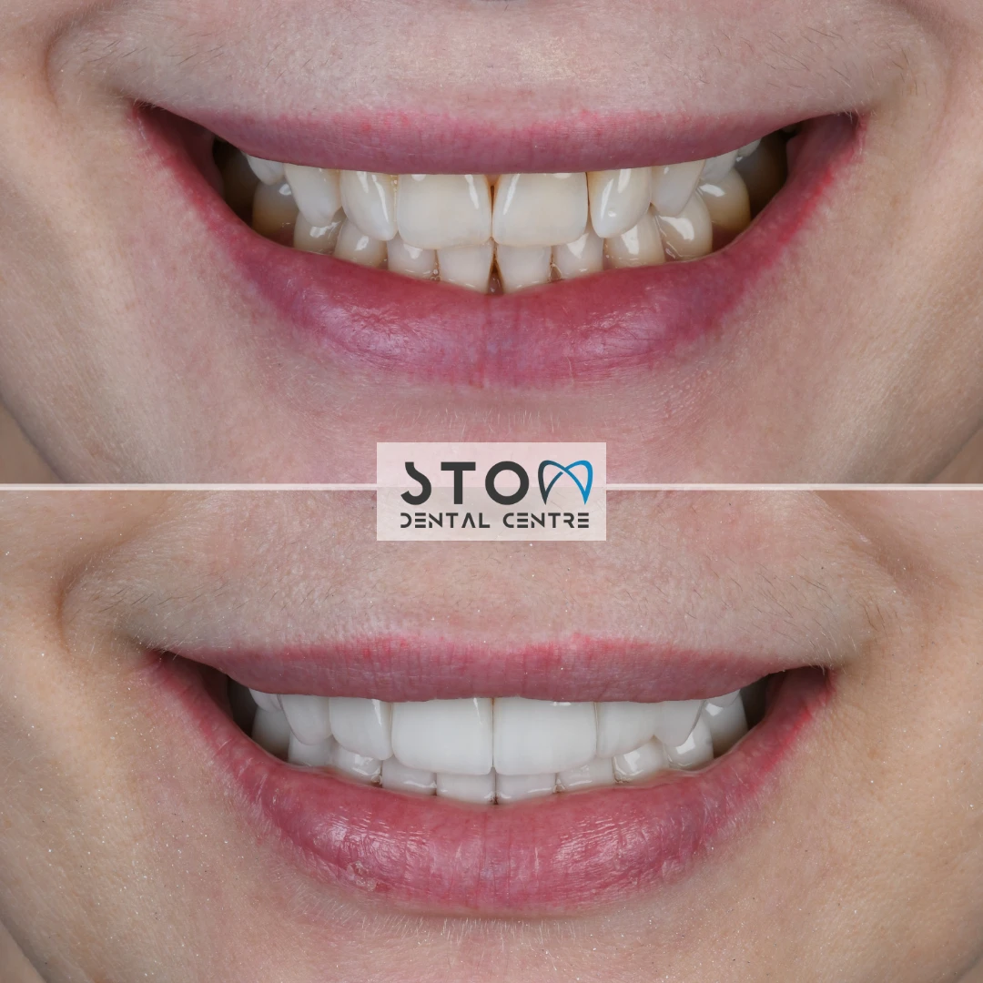
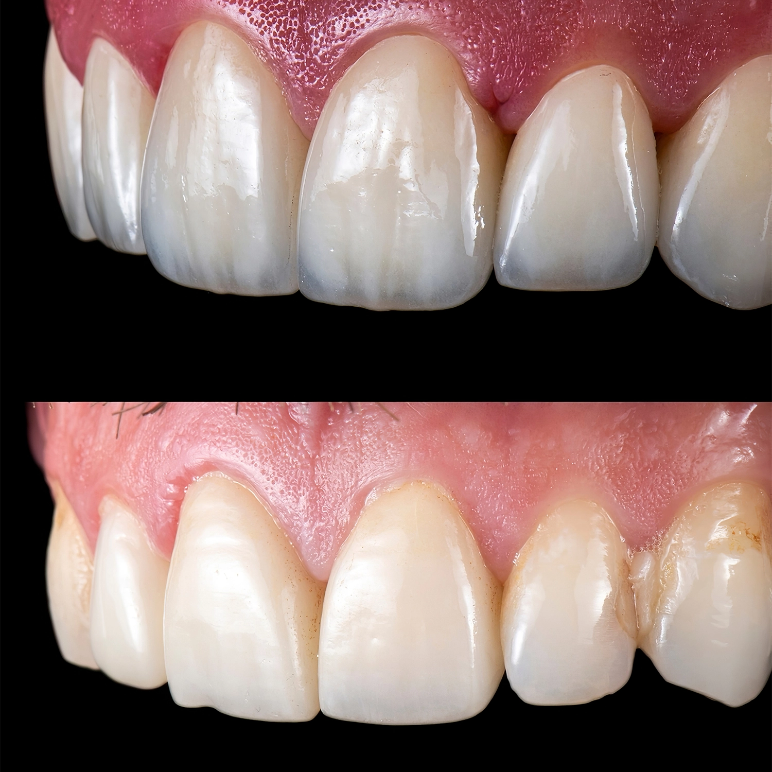
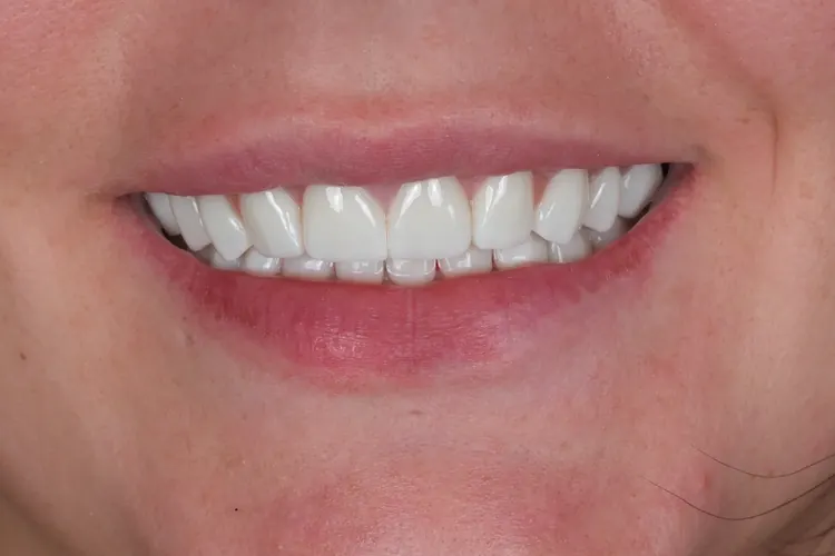
Before and after: patients of our clinic

 BeforeAfter

 BeforeAfter
 BeforeAfter
Photos taken at our clinic: cropped only, not retouched. Every result is individual.
See all before and after cases