What Is a CBCT Scan? 3D Dental X-Ray Explained — Radiation Dose, Uses, and When It’s Not Needed
Medically reviewed by Dr. Telman İskender, Clinical Director, Stom Dental Centre Antalya
Meet our team and patient coordinators — verified names and WhatsApp numbers, so you always know who you are talking to.
“Is the radiation safe, and do I actually need this?” — almost every patient asks some version of that question when a CBCT scan is recommended before an implant, a root canal, or a wisdom tooth extraction. The short answer: CBCT (Cone Beam Computed Tomography) is a 3D X-ray that shows the internal structure of your jaw in far more detail than a standard panoramic X-ray, at a radiation dose that is, in most cases, a fraction of a medical CT scan. Below is what it actually is, how its dose compares with real published numbers, and — just as importantly — when it is not needed at all.
- CBCT produces a 3D, sliced image of the jaw from a single rotation around the head, unlike a flat, two-dimensional panoramic X-ray.
- Dose depends on the scan area: small-FOV scans average ~84 µSv, large-FOV scans ~212 µSv — published values for a medical CT of the jaw region are roughly 430-1,410 µSv, i.e. several to about ten times higher depending on the device and scan area.
- For context: a small-FOV CBCT is roughly equivalent to about 13 days of natural background radiation, a large-FOV scan to about a month — a simple arithmetic comparison, not a claim of equal biological risk.
- Guided implant surgery based on CBCT deviates from the plan by an average of about 1.2 mm at the entry point and 1.4 mm at the apex — which is why a safety margin of at least 2 mm is recommended.
- CBCT should be used selectively, not routinely — the 2025 AAE/AAOMR joint position statement is explicit about this (ALADAIP principle).
What Exactly Is a CBCT Scan?
CBCT is an X-ray technique that produces a three-dimensional, sliced image of the jaw and teeth from a single rotation around the head. A conventional panoramic or periapical (single tooth) X-ray produces one flat, two-dimensional image; CBCT instead lets your dentist examine the same region layer by layer, from any angle — revealing bone density, the exact path of nerve canals, the number and shape of tooth roots, or hidden infections that simply don’t show up on a 2D film. Technically, it uses a cone-shaped X-ray beam (hence the name), which is why the equipment is far more compact and lower-dose than a hospital CT scanner (Scarfe & Farman, Dental Clinics of North America, 2008).

How Does CBCT Actually Work?
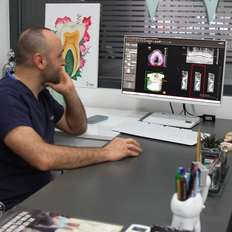
The scanner rotates once around your head — typically 180 to 360 degrees — while a cone-shaped X-ray beam and a flat-panel detector move together. Where a medical CT scanner takes dozens of separate thin slices, CBCT captures hundreds of images in a single rotation, and software reconstructs them into a 3D volume made of tiny cubes called voxels. The result is an image your dentist can rotate and section on screen from any angle. This design is both faster and, in most configurations, lower-dose than medical CT (Scarfe & Farman, 2008).

How Much Radiation Does CBCT Involve? (Compared to 2D X-Rays and Medical CT)
A comprehensive meta-analysis pooling data from nine different CBCT units found that effective dose depends heavily on the field of view (FOV) scanned: small-FOV scans (a single jaw segment, e.g. for one implant) average about 84 microsievert (µSv), with a range of 5-652 µSv; medium-FOV scans average 177 µSv; and large-FOV scans (both jaws) average 212 µSv, ranging 46-916 µSv (Ludlow et al., Dentomaxillofacial Radiology, 2015). For comparison: a single periapical X-ray delivers 0.65-9.5 µSv, a panoramic X-ray 3.85-38 µSv, while published values for a medical multi-slice CT of the jaw region range from about 430 to 1,410 µSv. In a study summarized in the same review, which defined the scanned area identically for both methods, CT doses were “several to ten times higher” than CBCT doses (Li, Imaging Science in Dentistry, 2013). At the extremes the ranges can overlap: some large-FOV CBCT protocols reach doses close to the lowest reported CT values, which is why the device and the scan area matter. For context, the worldwide average natural background radiation dose is about 2.4 millisievert per year, or roughly 6.6 µSv per day (UNSCEAR, 2008 Report). On that basis, a small-FOV CBCT scan is roughly equivalent to about 13 days of natural background radiation, and a large-FOV scan to about a month — a simple arithmetic comparison, not a claim of equivalent biological risk.
Comparison Table: 2D X-Ray vs. CBCT vs. Medical CT
| Method | Effective Dose (µSv) | Image Type | Typical Use | Source |
|---|---|---|---|---|
| Periapical (single tooth) X-ray | 0.65-9.5 | 2D, single tooth | Cavity checks, single-tooth follow-up | Li, 2013 |
| Panoramic X-ray | 3.85-38 | 2D, full-jaw overview | General assessment, orthodontic screening | Li, 2013 |
| CBCT (small FOV) | 5-652 (avg ~84) | 3D, limited region | Single implant, root canal detail | Ludlow et al., 2015 |
| CBCT (large FOV) | 46-916 (avg ~212) | 3D, both jaws | Multiple implants, jaw surgery planning | Ludlow et al., 2015 |
| Medical multi-slice CT (jaw region) | ~430-1,410 | 3D, hospital-grade, high resolution | Trauma, tumors, hospital settings | Li, 2013 |
What Is CBCT Used For in Dentistry?
Shows bone width and density, and the exact position of the inferior alveolar nerve canal in three dimensions — the basis for guided implant surgery.
Used selectively, per the 2025 AAE/AAOMR guidance, for extra canals, root fractures, complex anatomy, or cases that fail to heal.
Used when a panoramic X-ray suggests the tooth root is close to the nerve canal.
When a full 3D map of the bone is required.
To define the extent of a lesion and its relationship to surrounding structures.
What Does the Evidence Say About CBCT’s Advantages?
A systematic review and meta-analysis of guided implant surgery accuracy found that implants planned on CBCT-based surgical guides deviated from the planned position by an average of only about 1.2 mm at the entry point and 1.4 mm at the apex, with an average angular deviation of about 3.5 degrees (Tahmaseb et al., Clinical Oral Implants Research, 2018). That predictability lets the surgical team plan the distance to a nerve canal or an adjacent root in advance; because these are average values, the authors recommend respecting a safety margin of at least 2 mm. The AAOMR’s own position statement on implant imaging identifies cross-sectional (3D) imaging as the preferred method for assessing implant sites (Tyndall et al., Oral Surgery, Oral Medicine, Oral Pathology and Oral Radiology, 2012).

Does Every Patient Need a CBCT Scan? When It’s Not Necessary
No — and that honesty matters. The 2025 AAE/AAOMR joint position statement is explicit that CBCT “should not be used routinely but rather selectively,” guided by the ALADAIP principle (radiation As Low As Diagnostically Acceptable, Indication- and Patient-specific). A telling example: a systematic review of whether adding CBCT to panoramic imaging reduces nerve injury in wisdom tooth surgery found that only one of seven randomized controlled trials showed a significant benefit (4.2% vs 16.4% nerve injury, p=0.017); the other six found no significant difference between CBCT and panoramic imaging alone — meaning CBCT does not routinely lower nerve-injury risk outside genuinely high-risk cases (Robbins et al., BMC Oral Health, 2022). For a straightforward filling, a single-canal root treatment with normal anatomy, or a routine check-up, a panoramic or periapical X-ray is generally sufficient and CBCT is not indicated.
What Actually Happens During a CBCT Scan? (The Patient’s Experience)
There is nothing to be anxious about. You stand or sit in front of a machine that looks similar to a panoramic X-ray unit, rest your chin lightly on a support or bite on a small peg, and stay still for a few seconds while the scanner makes one rotation around your head. The actual scan takes roughly 10 to 40 seconds depending on the device, and pulsed-exposure technology on modern units often keeps the actual radiation-on time shorter still (Scarfe & Farman, 2008). No injection, no contrast dye, no special preparation is needed. Images are available digitally within seconds, and the whole appointment — including positioning — typically takes 10 to 15 minutes.
“A CBCT scan is a tool, not a routine step. The question I always ask first is whether the extra 3D detail will actually change the treatment plan — if it won’t, the patient doesn’t need the extra dose.”
How CBCT Fits Into Treatment at Stom Dental Centre
At Stom Dental Centre, CBCT is one of the diagnostic tools used for implant planning, complex root canal cases, wisdom tooth surgery, and Digital Smile Design (DSD) treatment planning. The decision to scan is made case by case, based on the clinical indication criteria outlined above — CBCT is not applied as a routine add-on to every treatment, but recommended where it is genuinely expected to change or improve the treatment plan.

Frequently Asked Questions
Is CBCT dangerous — does it cause cancer?
CBCT uses ionizing radiation and, in theory, carries a very small risk. Its dose is usually several times lower than that of a medical CT scan: depending on the device and scan area, studies report CT doses several to about ten times higher (Li, 2013). As simple arithmetic, a small-area scan corresponds on average to about two weeks and a large-area scan to about one month of natural background radiation (UNSCEAR, 2008). That’s why dentistry applies the ALADAIP principle — using it only when the diagnostic benefit justifies the dose.
Planning dental treatment in Antalya?
Get a free, no-obligation assessment from our clinical team — we reply within 24 hours.
How is CBCT different from a normal panoramic X-ray?
A panoramic X-ray gives one flat, two-dimensional image. CBCT produces a three-dimensional, sliced image that shows bone depth, nerve position, and root anatomy that a 2D film simply cannot capture (Scarfe & Farman, 2008).
How often can I have a CBCT scan?
There’s no fixed annual limit. Each scan is justified individually based on clinical need, and international guidance specifically warns against unnecessary repeat imaging (AAE & AAOMR, 2025).
Do I need a CBCT scan for every dental implant?
If guided implant surgery is planned, CBCT is strongly recommended — the AAOMR names cross-sectional imaging as the preferred method for implant site assessment (Tyndall et al., 2012). For simpler cases, your dentist decides based on the complexity of the site.
Can I have a CBCT scan while pregnant?
The general rule in dental radiography is that elective (non-urgent) imaging is postponed during pregnancy, or performed only if genuinely necessary and with appropriate shielding — this should always be discussed directly with your dentist.
Is CBCT the same as a medical CT scan?
No. Medical CT uses a higher dose, is also strong for imaging soft tissue, and is typically hospital-based. CBCT is optimized specifically for bone and dental structures, uses a lower dose, and is designed for use in a dental clinic (Li, 2013).
Does the scan hurt, and how long does it take?
No, it’s completely painless. The scan itself takes about 10 to 40 seconds depending on the device, and the full appointment, including positioning, is typically 10 to 15 minutes.
Do I always need a CBCT scan before wisdom tooth removal?
No — it’s recommended specifically when a panoramic X-ray suggests the tooth root is close to the nerve canal. A systematic review found that in lower-risk cases, adding CBCT does not routinely reduce the rate of nerve injury (Robbins et al., 2022).
Sources
- Scarfe WC, Farman AG. “What Is Cone Beam CT and How Does It Work?” Dental Clinics of North America. 2008;52(4):707-730.
- Ludlow JB, Timothy R, Walker C, Hunter R, Benavides E, Samuelson DB, Scheske MJ. “Effective dose of dental CBCT—a meta-analysis of published data and additional data for nine CBCT units.” Dentomaxillofacial Radiology. 2015;44(1):20140197.
- Li G. “Patient radiation dose and protection from cone-beam computed tomography.” Imaging Science in Dentistry. 2013;43(2):63-69.
- UNSCEAR (United Nations Scientific Committee on the Effects of Atomic Radiation). Sources and Effects of Ionizing Radiation, 2008 Report to the General Assembly — worldwide average natural background radiation ≈2.4 mSv/year.
- American Association of Endodontists (AAE) & American Academy of Oral and Maxillofacial Radiology (AAOMR). Joint Position Statement: “Use of Cone-Beam Computed Tomography in Endodontics — 2025 Update.” Journal of Endodontics. 2026;52(1):4-13.
- Robbins J, Smalley KR, Ray P, Ali K. “Does the addition of cone-beam CT to panoral imaging reduce inferior dental nerve injuries resulting from third molar surgery? A systematic review.” BMC Oral Health. 2022.
- Tahmaseb A, Wu V, Wismeijer D, Coucke W, Evans C. “The accuracy of static computer-aided implant surgery: A systematic review and meta-analysis.” Clinical Oral Implants Research. 2018;29(Suppl 16):416-435.
- Tyndall DA, Price JB, Tetradis S, Ganz SD, Hildebolt C, Scarfe WC; American Academy of Oral and Maxillofacial Radiology. “Position statement… on selection criteria for the use of radiology in dental implantology with emphasis on cone beam computed tomography.” Oral Surgery, Oral Medicine, Oral Pathology and Oral Radiology. 2012;113(6):817-826.
- Health Physics Society. “Radiation dose from dental cone-beam computed tomography systems (CBCT).” Ask the Experts, hps.org.
This article is for informational purposes only and does not replace an individual dental examination. The decision to perform a CBCT scan is always made by the treating dentist, based on each patient’s clinical situation, in line with the ALADAIP principle. Figures are drawn from independent scientific publications and are not a guarantee of outcome at any specific clinic.